Wilk Renata, Kluczewska Ewa, Syc Bożena, Bajor Grzegorz
Department of Anatomy, Medical University of Silesia, Katowice, Poland.
Pol J Radiol. 2011 Jul;76(3):16-25.
CURRENTLY, A FEW IMAGING METHODS ARE USED IN CNS DIAGNOSTICS: computed tomography - CT, magnetic resonance imaging - MRI, and ultrasonography - USG. The ventricular system changes its dimensions with child's development. Linear indices commonly used in the diagnostics of hydrocephalus do not consider developmental changes of the intracranial fluid spaces. The aim of our work was to identify reference values for selected linear indices in specific age groups.
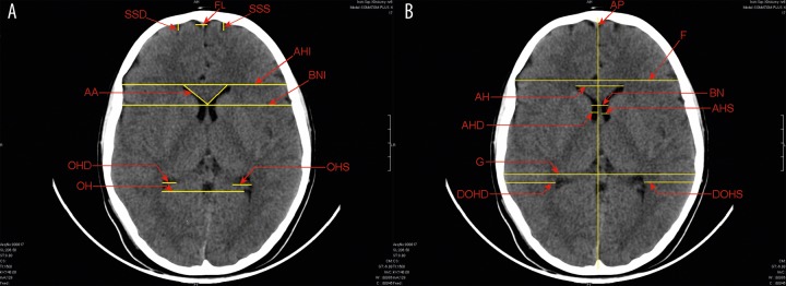
MATERIAL/METHODS: The material included 507 CT examinations of the head in children of different age and both sexes. There were 381 CT examinations considered as normal and they were used to establish the reference values. They were compared with 126 CTs from the observational zone (3-10 percentile and 90-97 percentile). The children were divided into 7 following age groups: 0-12 months, >12-36 months, >3-6 years, >6-9 years, >9-12 years, >12-15 years, >15-18 years. For every group, the 10(th), 25(th), 50(th), 75(th) and 90(th) percentile was calculated. The range between the 10(th) and the 90(th) percentile was described as a norm.
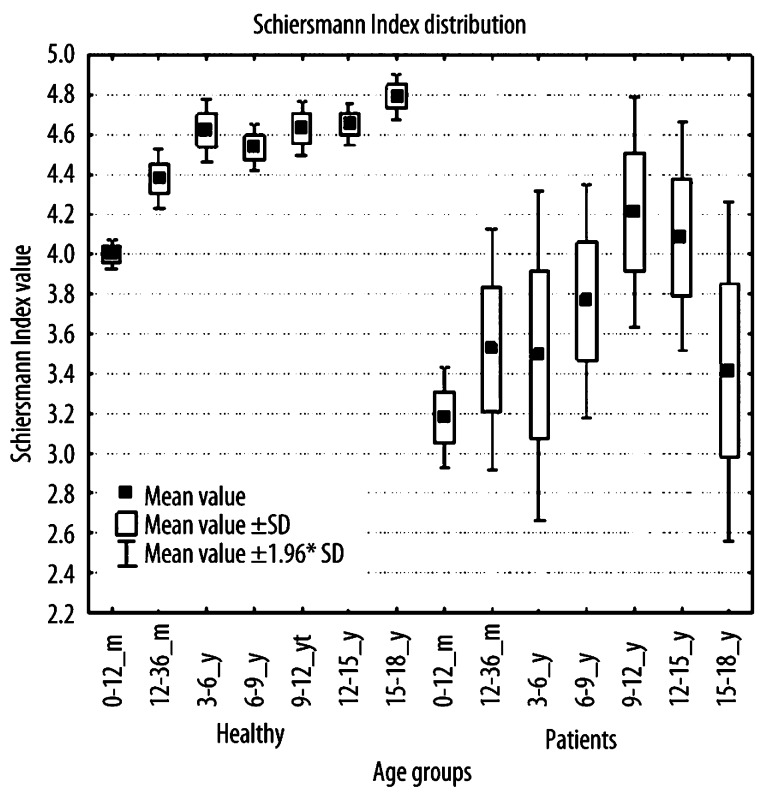
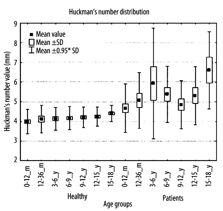
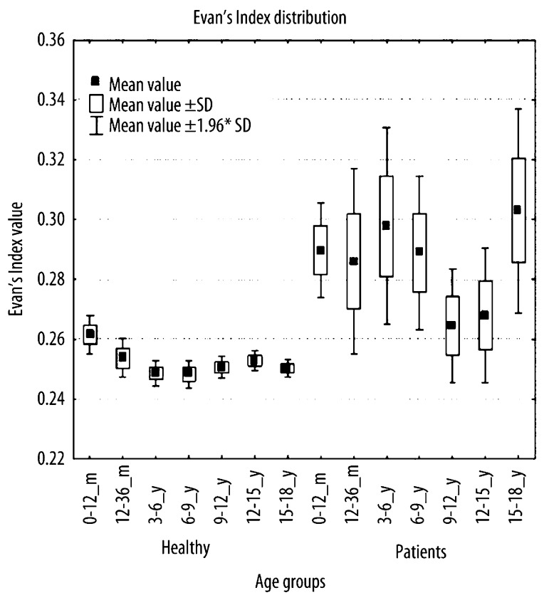
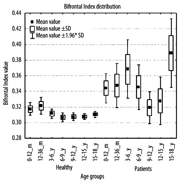
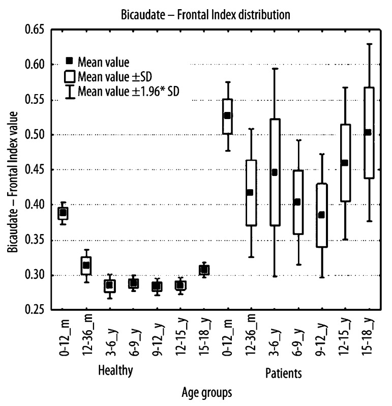
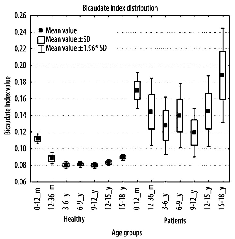
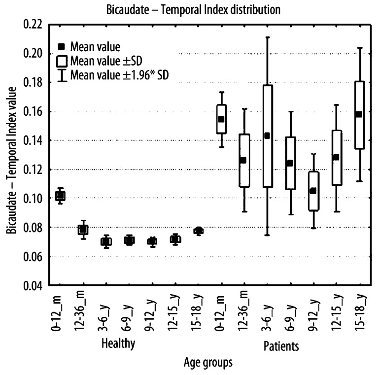
REFERENCE VALUES FOR PARTICULAR INDICES: Huckman Number from 3.3 to 5.0 cm with correlation coefficient according to age equal to 0.34; Evans' Index from 0.218 to 0.312 with correlation coefficient of -0.12; Bifrontal Index from 0.265 to 0.380 with correlation coefficient of 0.18; Bicaudate / Frontal Index from 0.212 to 0.524 with correlation coefficient of -0,33; Bicaudate Index from 0.059 to 0.152 with correlation coefficient of -0.26; Bicaudate / Temporal Index from 0.051 to 0.138 with correlation coefficient of 0.32; Schiersmann's Index from 3.545 to 6.038 with correlation coefficient of 0.42.
The intracerebral CSF spaces increased in a non-uniform manner with age. All indices established on the basis of linear parameters were relatively higher in younger children than in the older ones. In proportion to the cranial size, the intracranial fluid spaces decreased according to the age.
目前,中枢神经系统诊断中使用了几种成像方法:计算机断层扫描(CT)、磁共振成像(MRI)和超声检查(USG)。脑室系统会随着儿童的发育而改变其尺寸。脑积水诊断中常用的线性指标未考虑颅内液腔的发育变化。我们研究的目的是确定特定年龄组中选定线性指标的参考值。
材料/方法:材料包括507例不同年龄和性别的儿童头部CT检查。其中381例CT检查被视为正常,用于建立参考值。将它们与来自观察区的126例CT(第3至10百分位数和第90至97百分位数)进行比较。儿童被分为以下7个年龄组:0至12个月、大于12至36个月、大于3至6岁、大于6至9岁、大于9至12岁、大于12至15岁、大于15至18岁。对于每个组,计算第10、25、50、75和90百分位数。第10百分位数和第90百分位数之间的范围被描述为正常范围。
各指标的参考值:哈克曼数为3.3至5.0厘米,年龄相关系数为0.34;埃文斯指数为0.218至0.312,相关系数为-0.12;双额指数为0.265至0.380,相关系数为0.18;双尾状核/额指数为0.212至0.524,相关系数为-0.33;双尾状核指数为0.059至0.152,相关系数为-0.26;双尾状核/颞指数为0.051至0.138,相关系数为0.32;席尔斯曼指数为3.545至6.038,相关系数为0.42。
脑室内脑脊液间隙随年龄增长呈非均匀性增加。基于线性参数建立的所有指标在年幼儿童中相对高于年长儿童。与颅骨大小成比例,颅内液腔随年龄增长而减小。