Wolski Cyprian, Rotkiewicz Arkadiusz, Grzelak Piotr, Elgalal Marcin, Stefańczyk Ludomir
Department of Radiology and Diagnostic Imaging, Medical University of Łódź, Łódź, Poland.
Pol J Radiol. 2011 Oct;76(4):15-20.
At present, there is a number of diagnostic imaging procedures allowing for the evaluation of atherosclerosis. The earliest, subclinical stage of atherosclerosis can be visualized with the development of computed tomography (CT) and ultrasound (US) techniques. Therefore, the purpose of this study was to assess the degree of coronary artery calcification and carotid intima-media thickness in diabetic subjects divided into different age groups.
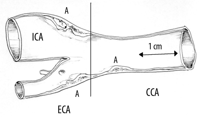
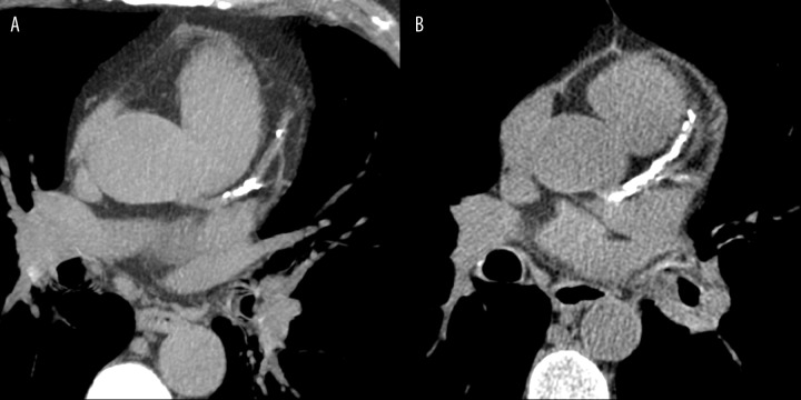
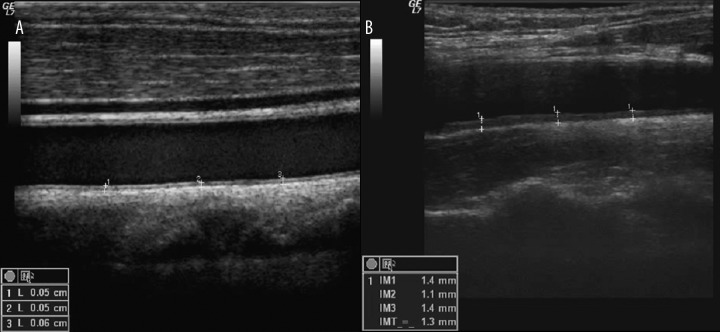
MATERIAL/METHODS: Fifty-six men, aged from 18 to 72 were included in the study. Participants were divided into 4 groups according to age (18-30, 31-45, 46-60 and more than 60 years). Two tests were performed: coronary calcium score (CS) determination and intima-media thickness (IMT) in ultrasound. CS was performed using a multi-slice scanner. Images were analyzed using the Agatson method. Ultrasound examinations were performed using a 9-12-MHz linear transducer.
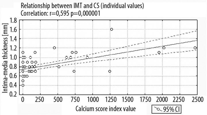
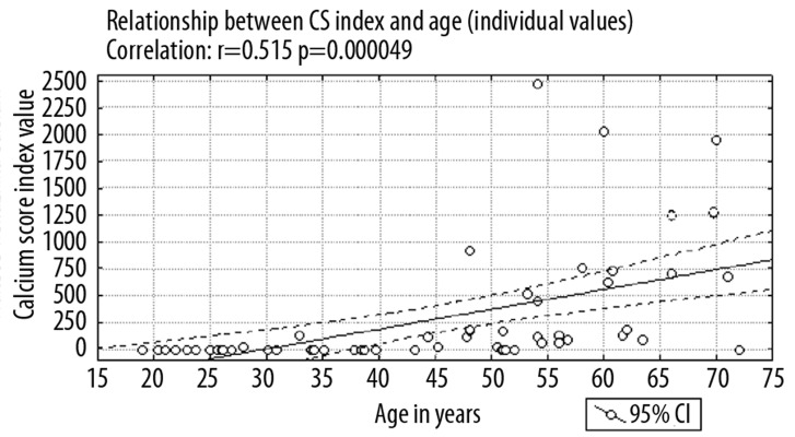
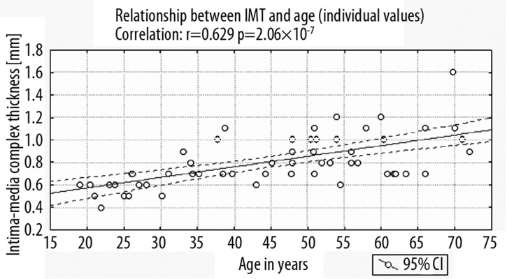
The correlation coefficient between calcium score index (CSI) and age of patients was 0.52 (p<0.001). The correlation between duration of diabetes and CSI was significantly lower (r=0.3; p<0.05). The increase of IMT is associated with age to a much greater extent and the correlation coefficient was 0.63 (p<0.001). IMT depended on the duration of diabetes, but the correlation was also weak (r=0.35; p<0.01).
Comparison of the findings obtained in the presented study and in the group of healthy subjects proves that influence of diabetes on vascular deterioration may be observed, even among young individuals. Obtained results allow to make the following conclusions: 1. Calcium score index remains low in the group of male patients with diabetes before the age of 45. 2. Intima-media thickness correlates well with age (r=0.6; p<0.05) and weaker with the duration of diabetes (r=0.35; p<0.05). 3. IMT assessment may be a useful tool to identify the increased predisposition to atherosclerosis, also before the age of 30.
目前,有多种诊断成像程序可用于评估动脉粥样硬化。随着计算机断层扫描(CT)和超声(US)技术的发展,动脉粥样硬化最早的亚临床阶段也能够被可视化。因此,本研究的目的是评估不同年龄组糖尿病患者的冠状动脉钙化程度和颈动脉内膜中层厚度。
材料/方法:本研究纳入了56名年龄在18至72岁之间的男性。参与者根据年龄分为4组(18 - 30岁、31 - 45岁、46 - 60岁和60岁以上)。进行了两项检查:冠状动脉钙化评分(CS)测定和超声内膜中层厚度(IMT)测量。CS使用多层扫描仪进行。图像采用阿加特森方法进行分析。超声检查使用9 - 12MHz线性换能器进行。
钙化评分指数(CSI)与患者年龄之间的相关系数为0.52(p<0.001)。糖尿病病程与CSI之间的相关性显著较低(r = 0.3;p<0.05)。IMT的增加与年龄的相关性更大,相关系数为0.63(p<0.001)。IMT取决于糖尿病病程,但相关性也较弱(r = 0.35;p<0.01)。
本研究结果与健康受试者组的比较证明,即使在年轻人中也可观察到糖尿病对血管恶化的影响。所得结果可得出以下结论:1. 45岁之前的男性糖尿病患者组的钙化评分指数仍然较低。2. 内膜中层厚度与年龄相关性良好(r = 0.6;p<0.05),与糖尿病病程的相关性较弱(r = 0.35;p<0.05)。3. IMT评估可能是一种有用的工具,可用于在30岁之前识别动脉粥样硬化易感性增加的情况。