Cardiovascular Research Center and Department of Physiology, Temple University School of Medicine, Philadelphia, Pennsylvania, United States of America.
PLoS One. 2012;7(7):e39965. doi: 10.1371/journal.pone.0039965. Epub 2012 Jul 13.
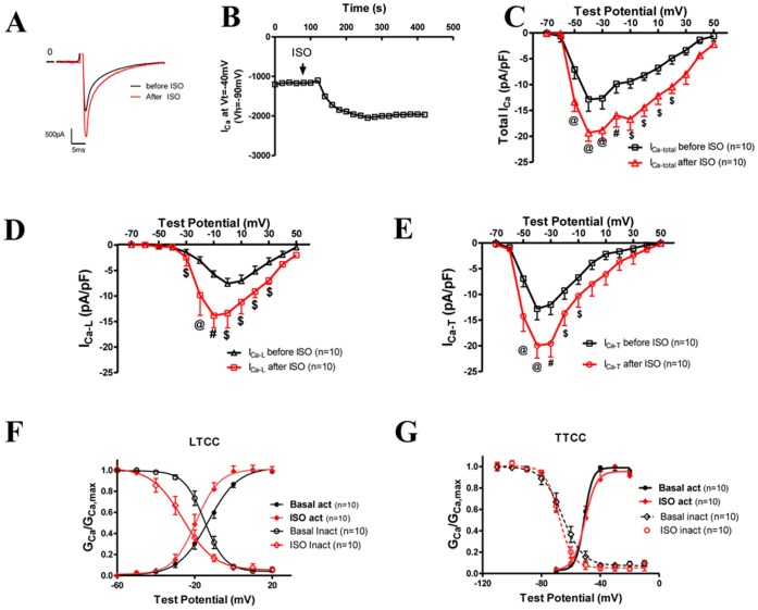
The T-type Ca(2+) channel (TTCC) plays important roles in cellular excitability and Ca(2+) regulation. In the heart, TTCC is found in the sinoatrial nodal (SAN) and conduction cells. Cav3.1 encodes one of the three types of TTCCs. To date, there is no report regarding the regulation of Cav3.1 by β-adrenergic agonists, which is the topic of this study. Ventricular myocytes (VMs) from Cav3.1 double transgenic (TG) mice and SAN cells from wild type, Cav3.1 knockout, or Cav3.2 knockout mice were used to study β-adrenergic regulation of overexpressed or native Cav3.1-mediated T-type Ca(2+) current (I(Ca-T(3.1))). I(Ca-T(3.1)) was not found in control VMs but was robust in all examined TG-VMs. A β-adrenergic agonist (isoproterenol, ISO) and a cyclic AMP analog (dibutyryl-cAMP) significantly increased I(Ca-T(3.1)) as well as I(Ca-L) in TG-VMs at both physiological and room temperatures. The ISO effect on I(Ca-L) and I(Ca-T) in TG myocytes was blocked by H89, a PKA inhibitor. I(Ca-T) was detected in control wildtype SAN cells but not in Cav3.1 knockout SAN cells, indicating the identity of I(Ca-T) in normal SAN cells is mediated by Cav3.1. Real-time PCR confirmed the presence of Cav3.1 mRNA but not mRNAs of Cav3.2 and Cav3.3 in the SAN. I(Ca-T) in SAN cells from wild type or Cav3.2 knockout mice was significantly increased by ISO, suggesting native Cav3.1 channels can be upregulated by the β-adrenergic (β-AR) system. In conclusion, β-adrenergic stimulation increases I(Ca-T(3.1)) in cardiomyocytes(,) which is mediated by the cAMP/PKA pathway. The upregulation of I(Ca-T(3.1)) by the β-adrenergic system could play important roles in cellular functions involving Cav3.1.
T 型钙通道(TTCC)在细胞兴奋和钙调节中发挥重要作用。在心脏中,TTCC 存在于窦房结(SAN)和传导细胞中。Cav3.1 编码三种 TTCC 之一。迄今为止,尚无关于β-肾上腺素能激动剂对 Cav3.1 的调节的报道,这是本研究的主题。使用 Cav3.1 双转基因(TG)小鼠的心室肌细胞(VM)和来自野生型、Cav3.1 敲除或 Cav3.2 敲除小鼠的 SAN 细胞来研究β-肾上腺素能对过表达或天然 Cav3.1 介导的 T 型钙电流(I(Ca-T(3.1)))的调节。在对照 VM 中未发现 I(Ca-T(3.1)),但在所有检查的 TG-VM 中均存在强烈的 I(Ca-T(3.1))。β-肾上腺素能激动剂(异丙肾上腺素,ISO)和环磷酸腺苷类似物(二丁酰环磷酸腺苷)在生理温度和室温下均显著增加 TG-VM 中的 I(Ca-T(3.1))和 I(Ca-L)。PKA 抑制剂 H89 阻断了 ISO 对 TG 心肌细胞中 I(Ca-L)和 I(Ca-T)的作用。在正常的 SAN 细胞中,I(Ca-T)在对照野生型 SAN 细胞中检测到,但在 Cav3.1 敲除的 SAN 细胞中未检测到,表明正常 SAN 细胞中 I(Ca-T)的身份是由 Cav3.1 介导的。实时 PCR 证实 SAN 中存在 Cav3.1 mRNA,但不存在 Cav3.2 和 Cav3.3 mRNA。ISO 显著增加了来自野生型或 Cav3.2 敲除小鼠的 SAN 细胞中的 I(Ca-T),表明天然 Cav3.1 通道可被β-肾上腺素能(β-AR)系统上调。总之,β-肾上腺素能刺激增加了心肌细胞中的 I(Ca-T(3.1)),这是由 cAMP/PKA 途径介导的。β-肾上腺素能系统对 I(Ca-T(3.1))的上调可能在涉及 Cav3.1 的细胞功能中发挥重要作用。