Department of Pediatrics, Yale University School of Medicine, New Haven, Connecticut, United States of America.
PLoS One. 2012;7(7):e40589. doi: 10.1371/journal.pone.0040589. Epub 2012 Jul 13.
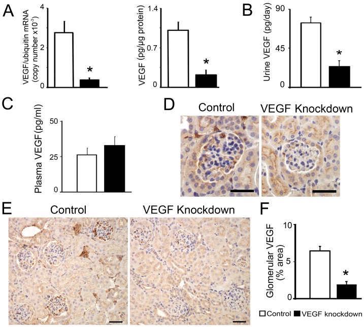
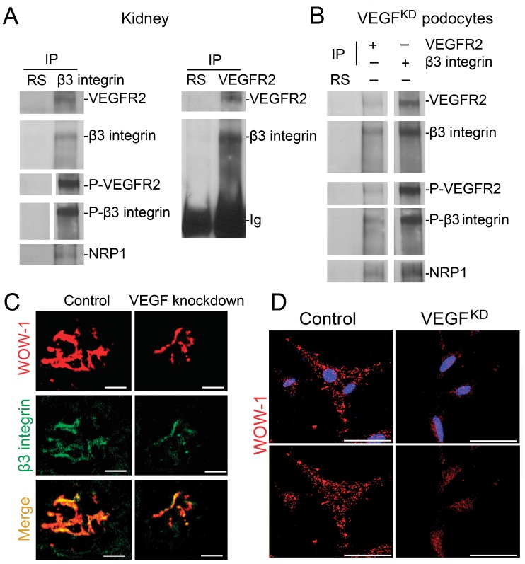
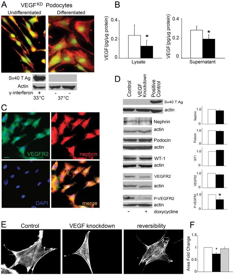
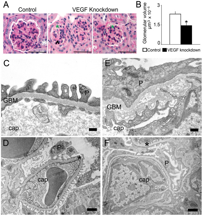
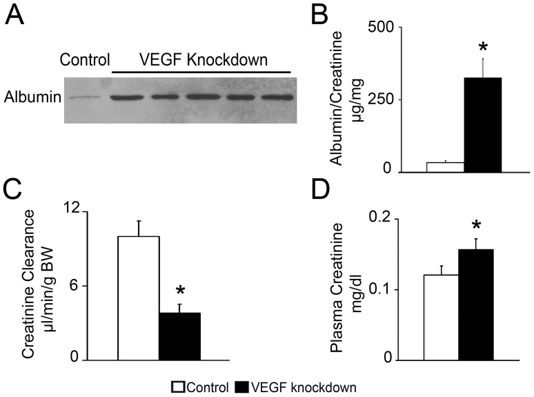
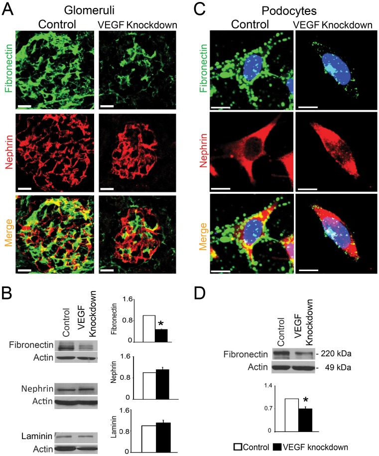
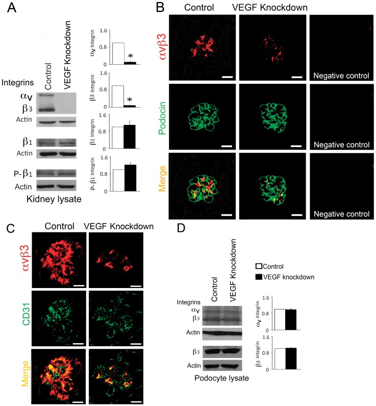
Podocyte or endothelial cell VEGF-A knockout causes thrombotic microangiopathy in adult mice. To study the mechanism involved in acute and local injury caused by low podocyte VEGF-A we developed an inducible, podocyte-specific VEGF-A knockdown mouse, and we generated an immortalized podocyte cell line (VEGF(KD)) that downregulates VEGF-A upon doxycycline exposure. Tet-O-siVEGF:podocin-rtTA mice express VEGF shRNA in podocytes in a doxycycline-regulated manner, decreasing VEGF-A mRNA and VEGF-A protein levels in isolated glomeruli to ~20% of non-induced controls and urine VEGF-A to ~30% of control values a week after doxycycline induction. Induced tet-O-siVEGF:podocin-rtTA mice developed acute renal failure and proteinuria, associated with mesangiolysis and microaneurisms. Glomerular ultrastructure revealed endothelial cell swelling, GBM lamination and podocyte effacement. VEGF knockdown decreased podocyte fibronectin and glomerular endothelial alpha(V)beta(3) integrin in vivo. VEGF receptor-2 (VEGFR2) interacts with beta(3) integrin and neuropilin-1 in the kidney in vivo and in VEGF(KD) podocytes. Podocyte VEGF knockdown disrupts alpha(V)beta(3) integrin activation in glomeruli, detected by WOW1-Fab. VEGF silencing in cultured VEGF(KD) podocytes downregulates fibronectin and disrupts alpha(V)beta(3) integrin activation cell-autonomously. Collectively, these studies indicate that podocyte VEGF-A regulates alpha(V)beta(3) integrin signaling in the glomerulus, and that podocyte VEGF knockdown disrupts alpha(V)beta(3) integrin activity via decreased VEGFR2 signaling, thereby damaging the three layers of the glomerular filtration barrier, causing proteinuria and acute renal failure.
足细胞或内皮细胞 VEGF-A 敲除可导致成年小鼠发生血栓性微血管病。为研究足细胞 VEGF-A 降低导致的急性和局部损伤的机制,我们构建了一种诱导型、足细胞特异性 VEGF-A 敲低小鼠,同时还构建了一种可诱导的永生化足细胞系(VEGF(KD)),该细胞系在给予强力霉素时可下调 VEGF-A 的表达。Tet-O-siVEGF:podocin-rtTA 小鼠以强力霉素调控的方式在足细胞中表达 VEGF shRNA,可使分离的肾小球中 VEGF-A mRNA 和 VEGF-A 蛋白水平降低至未诱导对照的 20%左右,尿液中 VEGF-A 水平降低至对照值的 30%左右,在强力霉素诱导 1 周后。诱导型 Tet-O-siVEGF:podocin-rtTA 小鼠发生急性肾衰竭和蛋白尿,伴有系膜溶解和微动脉瘤形成。肾小球超微结构显示内皮细胞肿胀、GBM 分层和足细胞足突消失。体内实验中 VEGF 敲低降低了足细胞纤维连接蛋白和肾小球内皮细胞 α(V)β(3)整联蛋白,体内和 VEGF(KD)足细胞中 VEGF 受体-2 (VEGFR2)与 β(3)整联蛋白和神经纤毛蛋白-1相互作用。足细胞 VEGF 敲低可破坏肾小球中 α(V)β(3)整联蛋白的激活,通过 WOW1-Fab 可检测到。在培养的 VEGF(KD)足细胞中沉默 VEGF 可下调纤维连接蛋白并破坏 α(V)β(3)整联蛋白的激活,这是通过 VEGFR2 信号的降低而实现的,从而破坏肾小球滤过屏障的三层结构,导致蛋白尿和急性肾衰竭。