XFEL Division; Japan Synchrotron Radiation Research Institute, Sayo-cho, Japan.
Nucleus. 2012 Sep-Oct;3(5):404-10. doi: 10.4161/nucl.21222. Epub 2012 Jul 31.
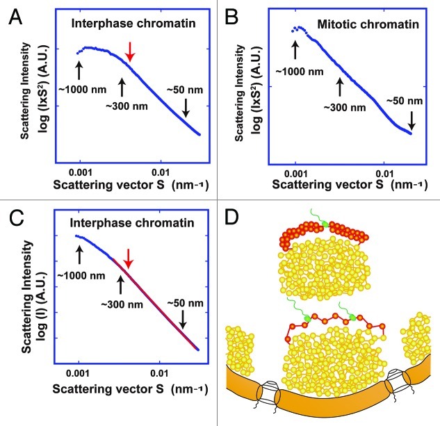
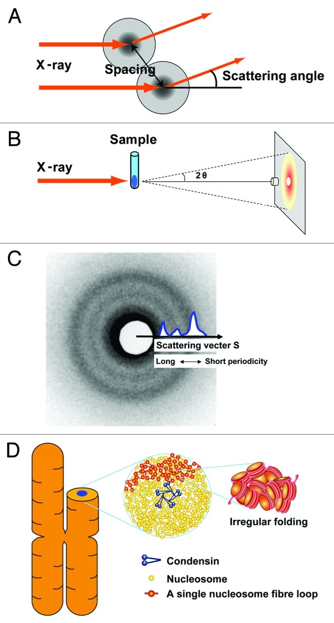
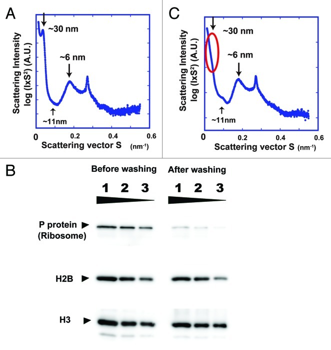
How is a long strand of genomic DNA packaged into a mitotic chromosome or nucleus? The nucleosome fiber (beads-on-a-string), in which DNA is wrapped around core histones, has long been assumed to be folded into a 30-nm chromatin fiber, and a further helically folded larger fiber. However, when frozen hydrated human mitotic cells were observed using cryoelectron microscopy, no higher-order structures that included 30-nm chromatin fibers were found. To investigate the bulk structure of mitotic chromosomes further, we performed small-angle X-ray scattering (SAXS), which can detect periodic structures in noncrystalline materials in solution. The results were striking: no structural feature larger than 11 nm was detected, even at a chromosome-diameter scale (~1 μm). We also found a similar scattering pattern in interphase nuclei of HeLa cells in the range up to ~275 nm. Our findings suggest a common structural feature in interphase and mitotic chromatins: compact and irregular folding of nucleosome fibers occurs without a 30-nm chromatin structure.
一条长的基因组 DNA 是如何包装成有丝分裂染色体或细胞核的?核小体纤维(珠串),其中 DNA 缠绕在核心组蛋白周围,长期以来一直被认为折叠成 30nm 的染色质纤维,以及进一步螺旋折叠的更大纤维。然而,当使用冷冻水合的人有丝分裂细胞进行冷冻电子显微镜观察时,没有发现包括 30nm 染色质纤维在内的更高阶结构。为了进一步研究有丝分裂染色体的整体结构,我们进行了小角度 X 射线散射(SAXS),它可以检测溶液中非晶态材料中的周期性结构。结果令人震惊:即使在染色体直径尺度(1μm)下,也没有检测到大于 11nm 的结构特征。我们还在 HeLa 细胞的间期核中发现了一个类似的散射模式,范围高达275nm。我们的发现表明,在有丝分裂和间期染色质中存在一个共同的结构特征:核小体纤维的紧密和不规则折叠,而没有 30nm 染色质结构。