Department of Radiation Physics, The University of Texas MD Anderson Cancer Center, Houston, TX, USA.
Radiat Oncol. 2012 Jul 24;7:116. doi: 10.1186/1748-717X-7-116.
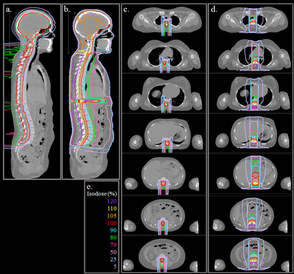
For many decades, the standard of care radiotherapy regimen for medulloblastoma has been photon (megavoltage x-rays) craniospinal irradiation (CSI). The late effects associated with CSI are well-documented in the literature and are in-part attributed to unwanted dose to healthy tissue. Recently, there is growing interest in using proton therapy for CSI in pediatric and adolescent patients to reduce this undesirable dose. Previous comparisons of dose to target and non-target organs from conventional photon CSI and passively scattered proton CSI have been limited to small populations (n ≤ 3) and have not considered the use of age-dependent target volumes in proton CSI.
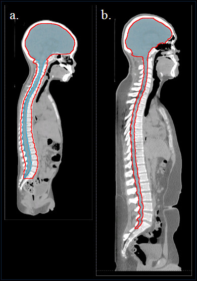
Standard of care treatment plans were developed for both photon and proton CSI for 18 patients. This cohort included both male and female medulloblastoma patients whose ages, heights, and weights spanned a clinically relevant and representative spectrum (age 2-16, BMI 16.4-37.9 kg/m2). Differences in plans were evaluated using Wilcoxon signed rank tests for various dosimetric parameters for the target volumes and normal tissue.
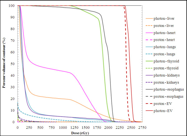
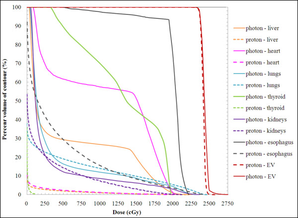
Proton CSI improved normal tissue sparing while also providing more homogeneous target coverage than photon CSI for patients across a wide age and BMI spectrum. Of the 24 parameters (V5, V10, V15, and V20 in the esophagus, heart, liver, thyroid, kidneys, and lungs) Wilcoxon signed rank test results indicated 20 were significantly higher for photon CSI compared to proton CSI (p ≤ 0.05) . Specifically, V15 and V20 in all six organs and V5, V10 in the esophagus, heart, liver, and thyroid were significantly higher with photon CSI.
Our patient cohort is the largest, to date, in which CSI with proton and photon therapies have been compared. This work adds to the body of literature that proton CSI reduces dose to normal tissue compared to photon CSI for pediatric patients who are at substantial risk for developing radiogenic late effects. Although the present study focused on medulloblastoma, our findings are generally applicable to other tumors that are treated with CSI.
几十年来,治疗髓母细胞瘤的标准治疗方案一直是光子(兆伏 X 射线)颅脊髓照射(CSI)。CSI 相关的晚期效应在文献中有详细记载,部分归因于对健康组织的不必要剂量。最近,人们对使用质子治疗小儿和青少年患者的 CSI 以减少这种不期望的剂量越来越感兴趣。以前对常规光子 CSI 和被动散射质子 CSI 的靶区和非靶区器官剂量的比较仅限于小人群(n≤3),并且没有考虑质子 CSI 中基于年龄的靶区体积的使用。
为 18 名患者制定了光子和质子 CSI 的标准治疗计划。该队列包括男性和女性髓母细胞瘤患者,其年龄、身高和体重涵盖了临床相关和代表性的范围(年龄 2-16 岁,BMI 16.4-37.9kg/m2)。使用 Wilcoxon 符号秩检验评估各种靶区和正常组织的剂量学参数的计划差异。
质子 CSI 改善了正常组织的保护,同时为广泛年龄和 BMI 范围内的患者提供了比光子 CSI 更均匀的靶区覆盖。在 24 个参数(食管、心脏、肝脏、甲状腺、肾脏和肺中的 V5、V10、V15 和 V20)中,Wilcoxon 符号秩检验结果表明,与质子 CSI 相比,光子 CSI 有 20 个参数显著更高(p≤0.05)。具体而言,所有六个器官的 V15 和 V20 以及食管、心脏、肝脏和甲状腺的 V5、V10 都有更高的光子 CSI。
我们的患者队列是迄今为止最大的,其中比较了质子和光子治疗的 CSI。这项工作增加了文献中的内容,即与光子 CSI 相比,质子 CSI 可减少小儿患者的正常组织剂量,这些患者有发生放射性晚期效应的高风险。尽管本研究侧重于髓母细胞瘤,但我们的发现通常适用于其他用 CSI 治疗的肿瘤。