Cellular and Molecular Metabolism Laboratory, Baker IDI Heart and Diabetes Institute, 75 Commercial Rd, Melbourne, VIC, 3004, Australia.
Department of Physiology, Monash University, Clayton, VIC, Australia.
Diabetologia. 2012 Oct;55(10):2769-2778. doi: 10.1007/s00125-012-2652-8. Epub 2012 Jul 26.
AIMS/HYPOTHESIS: Although skeletal muscle insulin resistance has been associated with activation of c-Jun N-terminal kinase (JNK), whether increased JNK activity causes insulin resistance in this organ is not clear. In this study we examined the metabolic consequences of isolated JNK phosphorylation in muscle tissue.
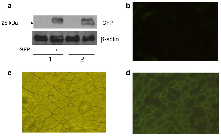
Plasmids containing genes encoding a wild-type JNK1 (WT-JNK) or a JNK1/JNKK2 fusion protein (rendering JNK constitutively active; CA-Jnk) were electroporated into one tibialis anterior (TA) muscle of C57Bl/6 mice, with the contralateral TA injected with an empty vector (CON) to serve as a within-animal control.
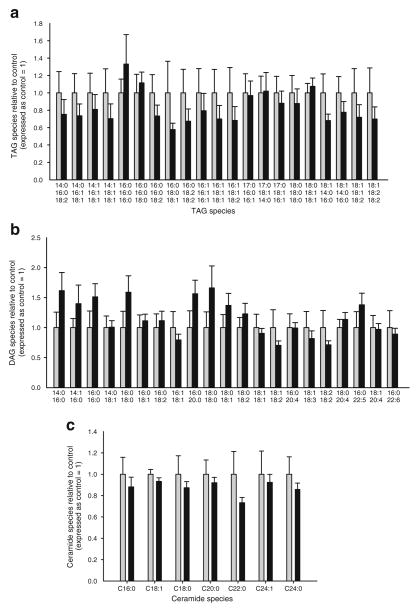
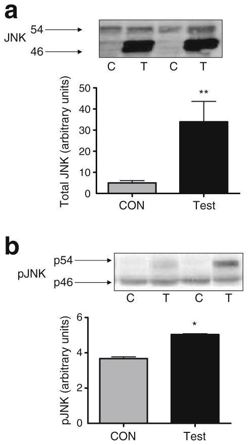
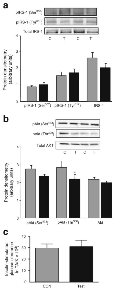
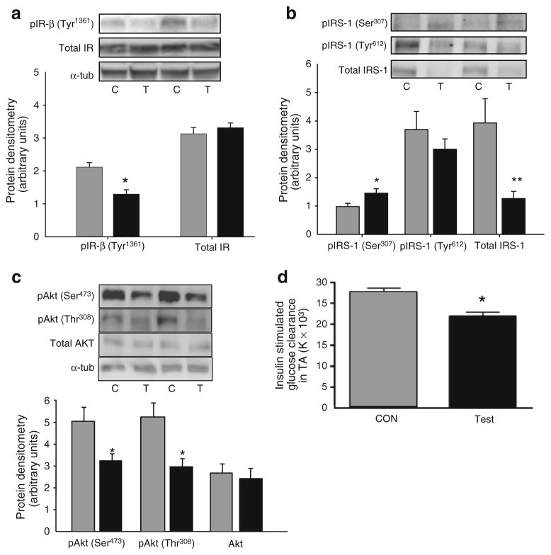
Overproduction of WT-JNK resulted in a modest (~25%) increase in phosphorylation (Thr(183)/Tyr(185)) of JNK, but no differences were observed in Ser(307) phosphorylation of insulin receptor substrate 1 (IRS-1) or total IRS-1 protein, nor in insulin-stimulated glucose clearance into the TA muscle when comparing WT-JNK with CON. By contrast, overexpression of CA-Jnk, which markedly increased the phosphorylation of CA-JNK, also increased serine phosphorylation of IRS-1, markedly decreased total IRS-1 protein, and decreased insulin-stimulated phosphorylation of the insulin receptor (Tyr(1361)) and phosphorylation of Akt at (Ser(473) and Thr(308)) compared with CON. Moreover, overexpression of CA-Jnk decreased insulin-stimulated glucose clearance into the TA muscle compared with CON and these effects were observed without changes in intramuscular lipid species.
CONCLUSIONS/INTERPRETATION: Constitutive activation of JNK in skeletal muscle impairs insulin signalling at the level of IRS-1 and Akt, a process which results in the disruption of normal glucose clearance into the muscle.
目的/假设:尽管骨骼肌胰岛素抵抗与 c-Jun N 末端激酶(JNK)的激活有关,但增加的 JNK 活性是否会导致该器官的胰岛素抵抗尚不清楚。在这项研究中,我们研究了肌肉组织中 JNK 磷酸化的代谢后果。
将含有编码野生型 JNK1(WT-JNK)或 JNK1/JNKK2 融合蛋白(使 JNK 持续激活;CA-Jnk)的基因的质粒电穿孔到 C57Bl/6 小鼠的一条比目鱼肌(TA)中,将另一条 TA 注射空载体(CON)作为体内对照。
WT-JNK 的过度表达导致 JNK 的磷酸化(Thr(183)/Tyr(185))适度增加(~25%),但在胰岛素受体底物 1(IRS-1)的 Ser(307)磷酸化或总 IRS-1 蛋白方面没有差异,也没有观察到 WT-JNK 与 CON 相比,胰岛素刺激 TA 肌肉葡萄糖清除率的差异。相比之下,CA-JNK 的过度表达显著增加了 CA-JNK 的磷酸化,也增加了 IRS-1 的丝氨酸磷酸化,显著降低了总 IRS-1 蛋白,并降低了胰岛素刺激的胰岛素受体(Tyr(1361))和 Akt 的磷酸化(Ser(473)和 Thr(308))与 CON 相比。此外,与 CON 相比,CA-JNK 的过度表达降低了胰岛素刺激 TA 肌肉的葡萄糖清除率,并且这些作用是在肌肉内脂质种类没有变化的情况下观察到的。
结论/解释:JNK 在骨骼肌中的组成性激活会损害 IRS-1 和 Akt 水平的胰岛素信号转导,从而导致正常的葡萄糖清除到肌肉中受到破坏。