Choromańska Agnieszka, Macura Katarzyna J
Department of Radiology, Military Institute of Medicine, Warsaw, Poland.
Pol J Radiol. 2012 Apr;77(2):22-34. doi: 10.12659/pjr.882967.
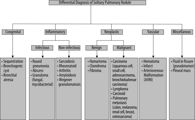
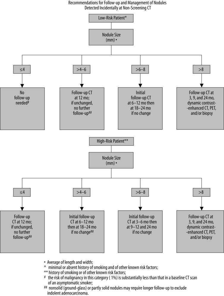
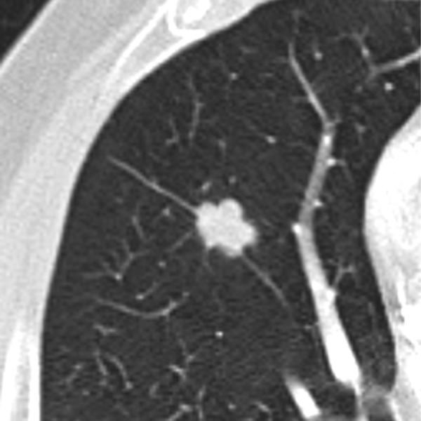
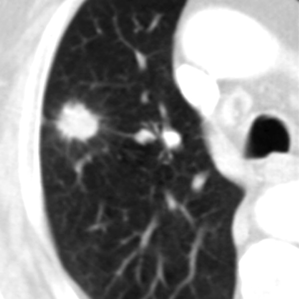
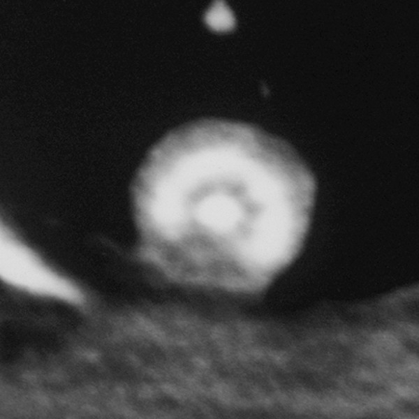
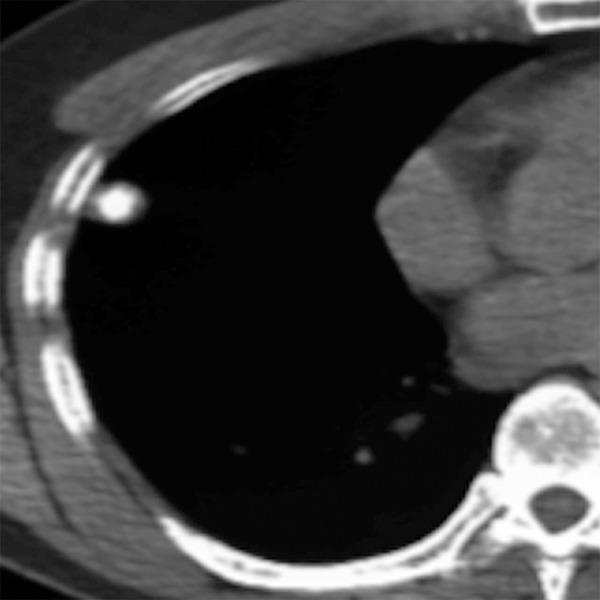
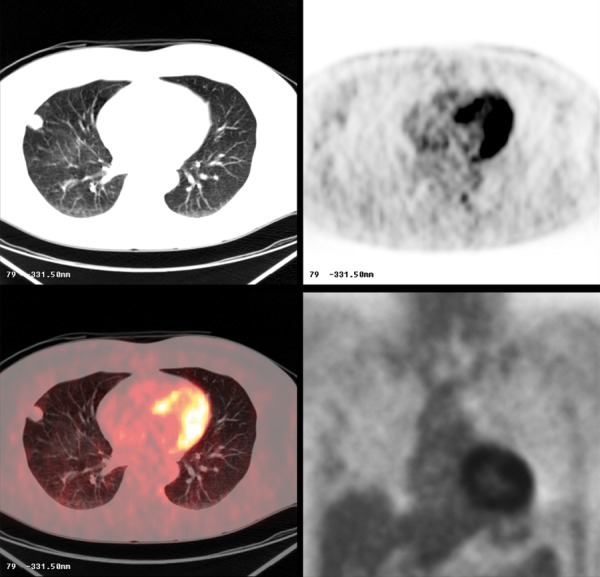
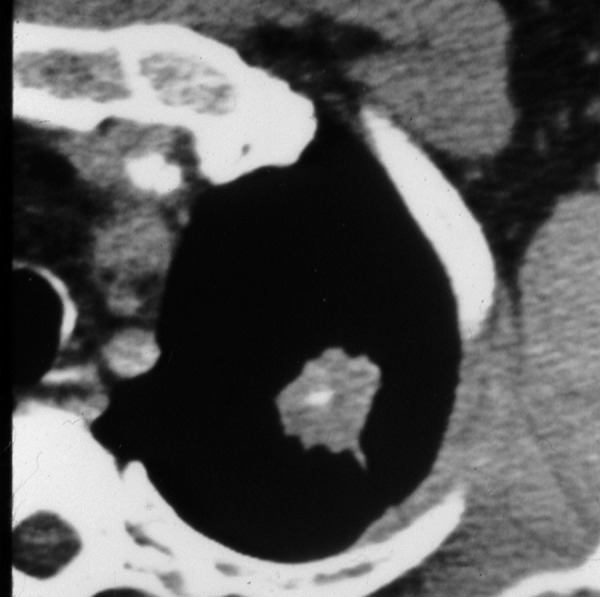
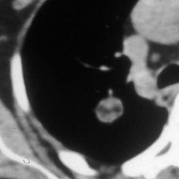
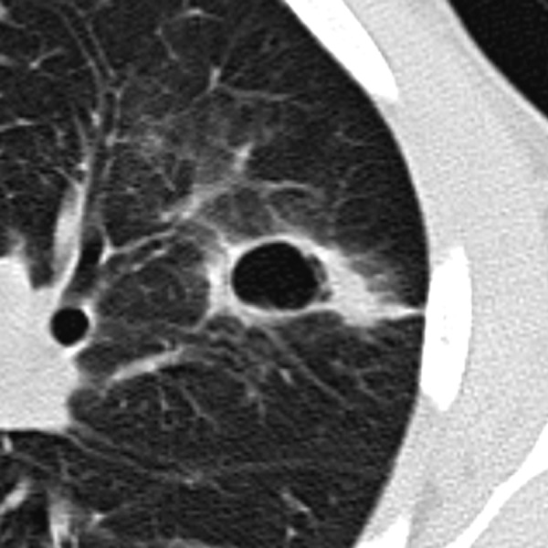
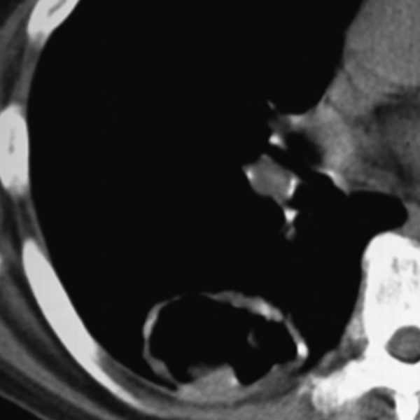
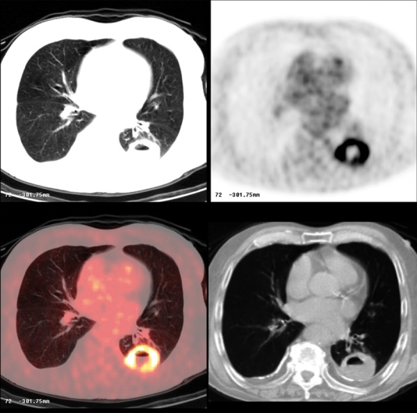
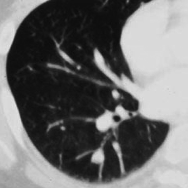
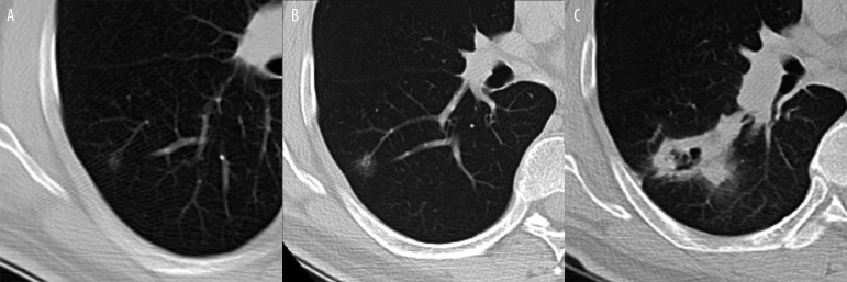
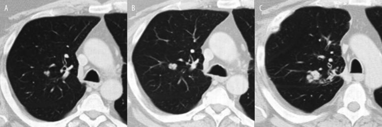
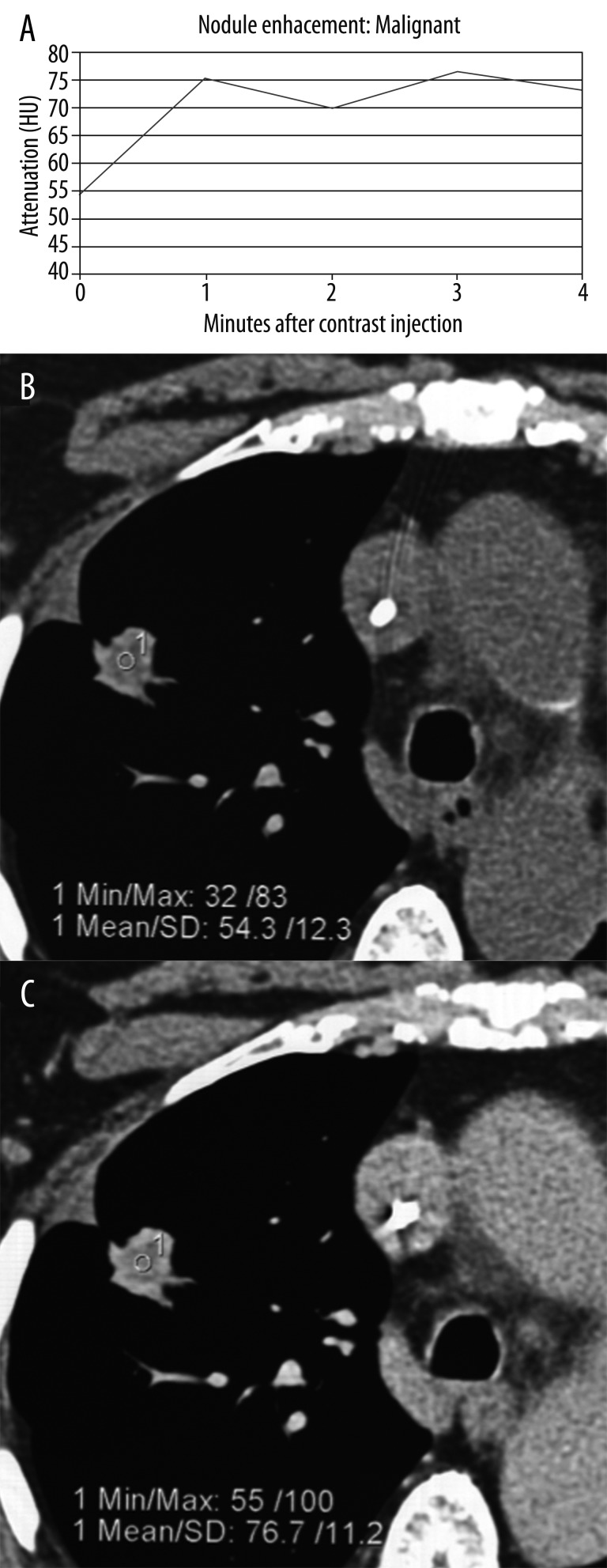
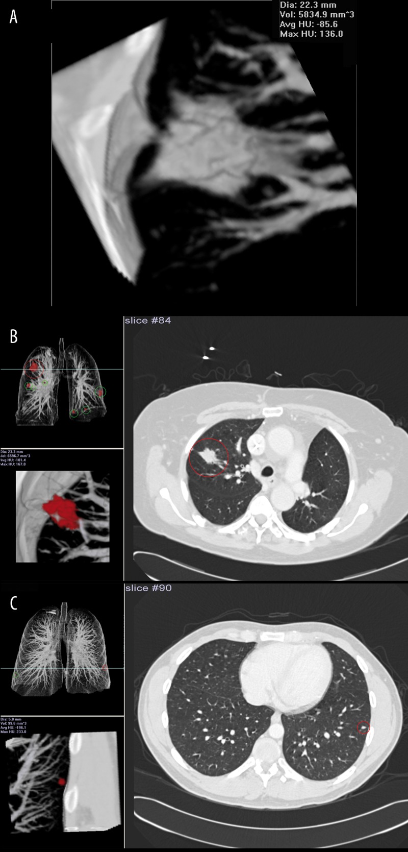
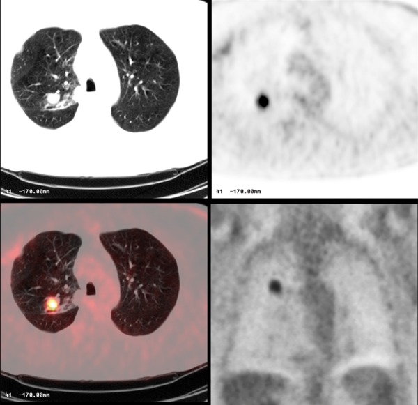
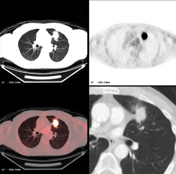
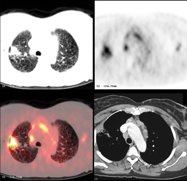
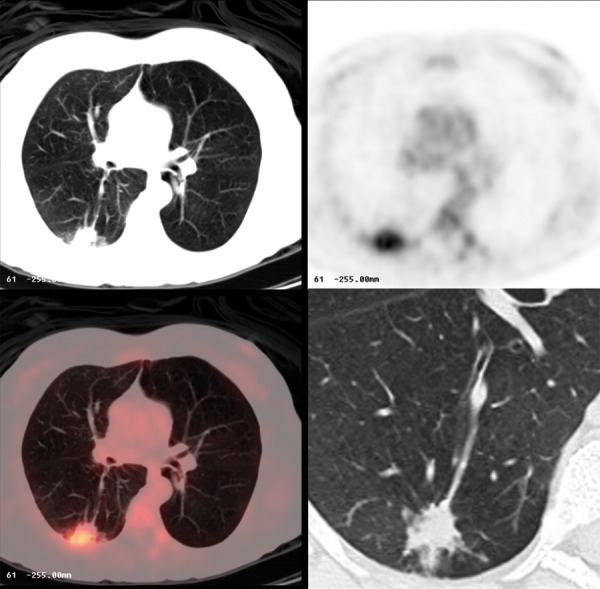
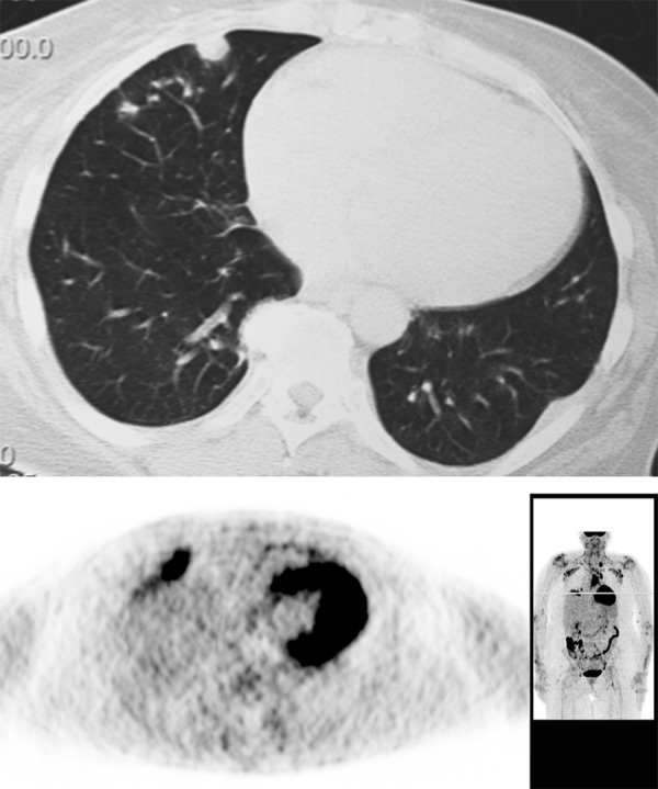
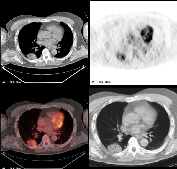
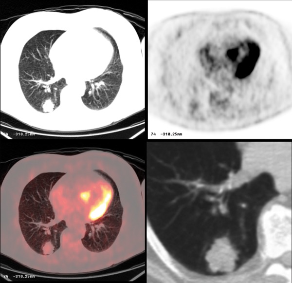
The solitary pulmonary nodule (SPN) has always been a diagnostic challenge for the radiologists. Currently, with increased utilization of computed tomography (CT) greater number of nodules is being discovered, with numerous indeterminate lesions, which frequently cannot be immediately classified into benign or malignant category.In this article we review the imaging features of benign and malignant round opacities; we demonstrate currently used standards and also more advanced techniques that are helpful in evaluating SPNs such as contrast-enhanced CT, PET/CT imaging and also pathologic sampling with biopsy or surgical resection.We also summarize the methods of evaluating and managing SPNs based on the latest guidelines from the Fleischner Society and American College of Chest Physicians.
孤立性肺结节(SPN)一直是放射科医生面临的诊断挑战。目前,随着计算机断层扫描(CT)的使用增加,发现了更多的结节,其中有许多不确定病变,这些病变常常无法立即归类为良性或恶性。在本文中,我们回顾了良性和恶性圆形致密影的影像学特征;我们展示了目前使用的标准以及更先进的技术,这些技术有助于评估SPN,如对比增强CT、PET/CT成像以及通过活检或手术切除进行病理采样。我们还根据弗莱施纳学会和美国胸科医师学会的最新指南总结了评估和处理SPN的方法。