Key Laboratory of Animal Reproduction and Germplasm Enhancement in Universities of Shandong, Qingdao Agricultural University, Qingdao, China.
PLoS One. 2012;7(7):e41771. doi: 10.1371/journal.pone.0041771. Epub 2012 Jul 24.
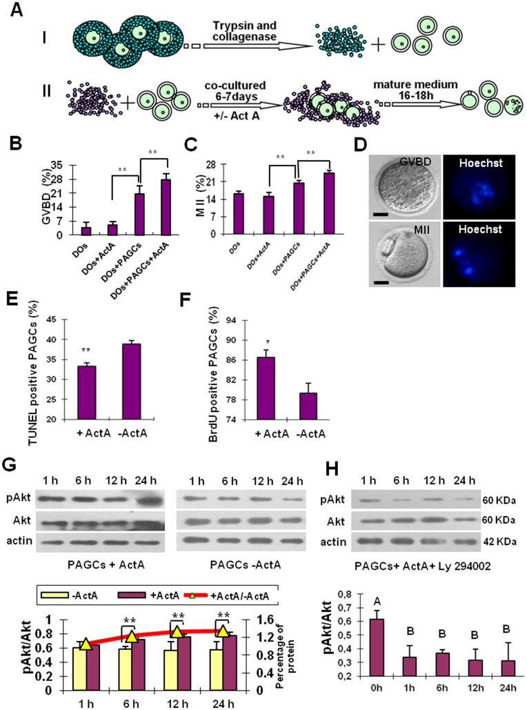
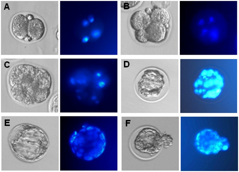
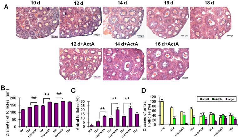
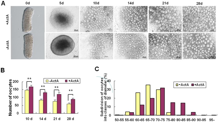
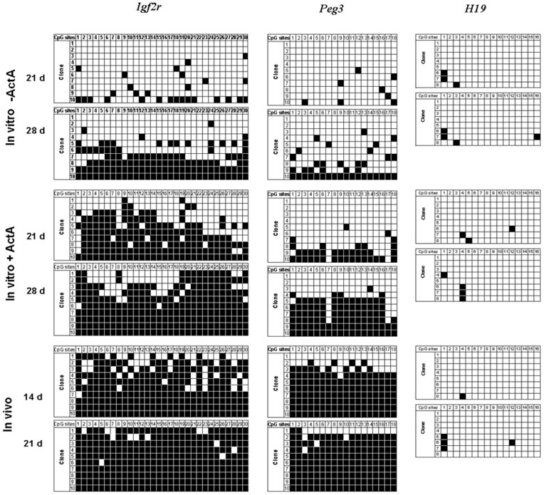
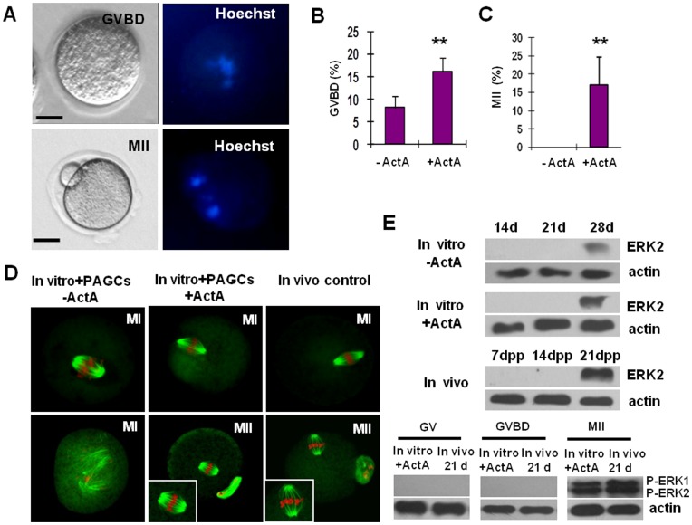
In the present study, we established an in vitro culture system suitable for generating fertilizable oocytes from premeiotic mouse female germ cells. These results were achieved after first establishing an in vitro culture system allowing immature oocytes from 12-14 day-old mice to reach meiotic maturation through culture onto preantral granulosa cell (PAGC) monolayers in the presence of Activin A (ActA). To generate mature oocytes from premeiotic germ cells, pieces of ovaries from 12.5 days post coitum (dpc) embryos were cultured in medium supplemented with ActA for 28 days and the oocytes formed within the explants were isolated and cocultured onto PAGC monolayers in the presence of ActA for 6-7 days. The oocytes were then subjected to a final meiotic maturation assay to evaluate their capability to undergo germinal vesicle break down (GVBD) and reach the metaphase II (MII) stage. We found that during the first 28 days of culture, a significant number of oocytes within the ovarian explants reached nearly full growth and formed preantral follicle-like structures with the surrounding somatic cells. GSH level and Cx37 expression in the oocytes within the explants were indicative of proper developmental conditions. Moreover, the imprinting of Igf2r and Peg3 genes in these oocytes was correctly established. Further culture onto PAGCs in the presence of ActA allowed about 16% of the oocytes to undergo GVBD, among which 17% reached the MII stage during the final 16-18 hr maturation culture. These MII oocytes showed normal spindle and chromosome assembly and a correct ERK1/2 activity. About 35% of the in vitro matured oocytes were fertilized and 53.44% of them were able to reach the 2-cell stage. Finally, around 7% of the 2-cell embryos developed to the morula/blastocyst stage.
在本研究中,我们建立了一种体外培养体系,能够从小鼠前体生殖细胞中生成可受精的卵母细胞。这些结果是通过首先建立一种体外培养体系实现的,该体系允许来自 12-14 天大的小鼠的不成熟卵母细胞在激活素 A(ActA)存在下培养到前腔颗粒细胞(PAGC)单层上,从而达到减数分裂成熟。为了从小鼠前体生殖细胞中生成成熟卵母细胞,将来自 12.5 天合胞体(dpc)胚胎的卵巢组织块在添加 ActA 的培养基中培养 28 天,然后分离出在培养物中形成的卵母细胞,并在添加 ActA 的情况下将其共培养到 PAGC 单层上 6-7 天。然后,将这些卵母细胞进行最后的减数分裂成熟测定,以评估它们进行生发泡破裂(GVBD)并达到中期 II(MII)阶段的能力。我们发现,在培养的前 28 天,卵巢培养物中的大量卵母细胞达到了几乎完全的生长,并形成了具有周围体细胞的前腔卵泡样结构。卵母细胞内 GSH 水平和 Cx37 表达表明发育条件良好。此外,这些卵母细胞中 Igf2r 和 Peg3 基因的印迹正确建立。进一步在 ActA 存在下培养到 PAGC 上,使大约 16%的卵母细胞发生 GVBD,其中 17%在最后的 16-18 小时成熟培养中达到 MII 阶段。这些 MII 卵母细胞显示出正常的纺锤体和染色体组装以及正确的 ERK1/2 活性。约 35%的体外成熟卵母细胞受精,其中 53.44%能够达到 2 细胞阶段。最后,约 7%的 2 细胞胚胎发育到桑椹胚/囊胚阶段。