Department of Biochemistry and Molecular Biology, and ARC Centre of Excellence in Structural and Functional Microbial Genomics, Monash University, Clayton, Victoria, Australia.
PLoS One. 2012;7(7):e42298. doi: 10.1371/journal.pone.0042298. Epub 2012 Jul 27.
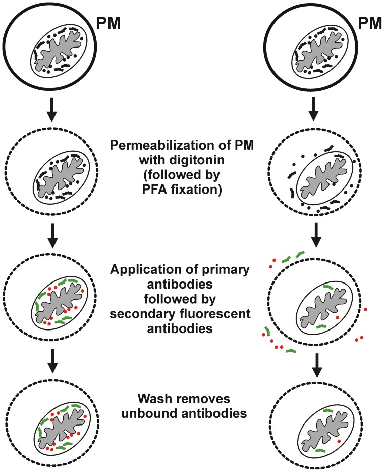
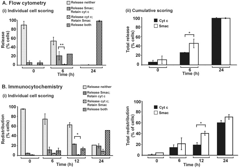
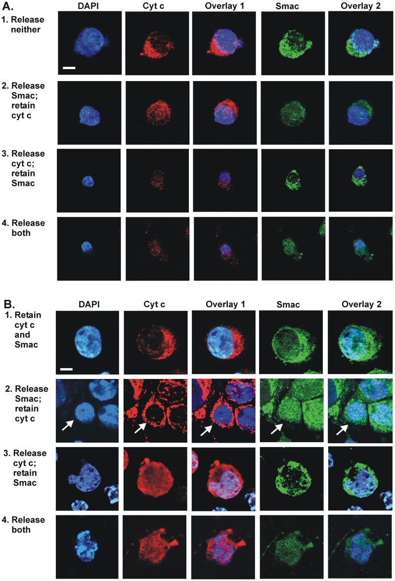
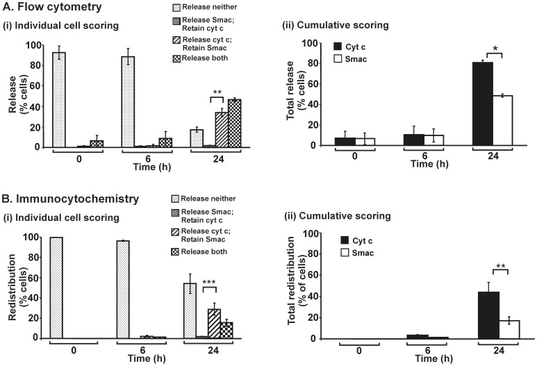
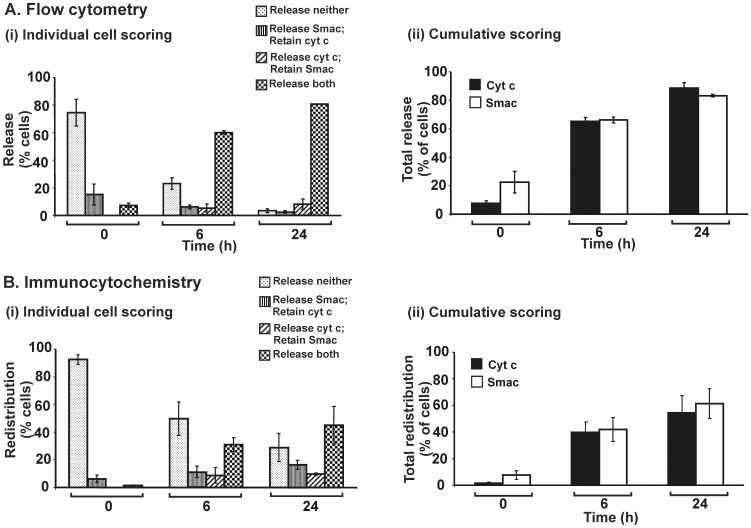
Mitochondrially mediated apoptosis is characterized by redistribution of proteins from mitochondria to cytoplasm following permeabilization of the outer mitochondrial membrane. We applied flow cytometry to quantify simultaneously the redistribution of two apoptogenic proteins, cytochrome c (cyt c) and Smac/DIABLO (Smac). Mammalian cells were treated with digitonin that selectively permeabilizes the plasma membrane. Following fixation, treated cells were infused successively with primary and secondary antibodies (the latter fluorescently tagged) enabling independent detection of cyt c and Smac. Digitonin-treated cells that retain cyt c or Smac in mitochondria generate strong fluorescence signals in flow cytometry. Cells in which cyt c or Smac have transited the outer mitochondrial membrane show greatly reduced fluorescence because the proteins are lost from the digitonin-permeabilized cells. Quantitative flow cytometry revealed that in 143B TK(-) cells treated with staurosporine, cyt c and Smac exit mitochondria asymmetrically, with cyt c redistribution preceding that of Smac. However, in HeLa cells likewise treated, cyt c and Smac exit mitochondria concurrently. Under other conditions of apoptotic induction, for example, 143B TK(-) cells treated with MT-21 (an apoptotic inducer that binds to the mitochondrial adenine nucleotide transporter), redistribution of Smac precedes that of cyt c. The various patterns of redistribution of these proteins were confirmed by immunocytochemical analysis and confocal microscopy. We conclude that flow cytometry can be employed effectively to quantify simultaneously the redistribution of cyt c and Smac from mitochondria to the cytosol. Moreover, differential redistribution of cyt c and Smac occurs under various conditions, thereby reflecting constraints on availability of these proteins to exit mitochondria after permeabilization of the outer membrane.
线粒体介导的细胞凋亡的特征是,在外膜通透性增加后,线粒体蛋白向细胞质重新分布。我们采用流式细胞术来定量分析两种促凋亡蛋白细胞色素 c(cyt c)和 Smac/DIABLO(Smac)的重新分布。用去污剂(如二丁基琥珀酸酯)处理哺乳动物细胞,该去污剂选择性地破坏质膜的通透性。处理后,细胞固定,然后依次用一抗和二抗(后者为荧光标记)孵育,以实现 cyt c 和 Smac 的独立检测。保留 cyt c 或 Smac 的二丁基琥珀酸酯处理细胞在流式细胞术中会产生强烈的荧光信号。而 cyt c 或 Smac 已穿过外膜的细胞,由于蛋白从二丁基琥珀酸酯处理的细胞中丢失,荧光大大降低。定量流式细胞术显示,在用 staurosporine 处理的 143B TK(-)细胞中,cyt c 和 Smac 不对称地从线粒体中释放,cyt c 的重新分布先于 Smac。然而,在同样用 staurosporine 处理的 HeLa 细胞中,cyt c 和 Smac 则同时从线粒体中释放。在其他诱导凋亡的条件下,例如用 MT-21(一种与线粒体腺嘌呤核苷酸转运蛋白结合的凋亡诱导剂)处理的 143B TK(-)细胞中,Smac 的重新分布先于 cyt c。这些蛋白的各种重新分布模式通过免疫细胞化学分析和共聚焦显微镜得到了证实。我们得出结论,流式细胞术可有效地用于定量分析 cyt c 和 Smac 从线粒体向细胞质的重新分布。此外,在不同条件下,cyt c 和 Smac 的重新分布存在差异,这反映了在外膜通透性增加后,这些蛋白从线粒体释放的限制。