Li Wen-Fei, Sun Ying, Chen Mo, Tang Ling-Long, Liu Li-Zhi, Mao Yan-Ping, Chen Lei, Zhou Guan-Qun, Li Li, Ma Jun
State Key Laboratory of Oncology in South China, Guangzhou, Guangdong 510060, P. R. China.
Chin J Cancer. 2012 Dec;31(12):579-87. doi: 10.5732/cjc.012.10095. Epub 2012 Aug 2.
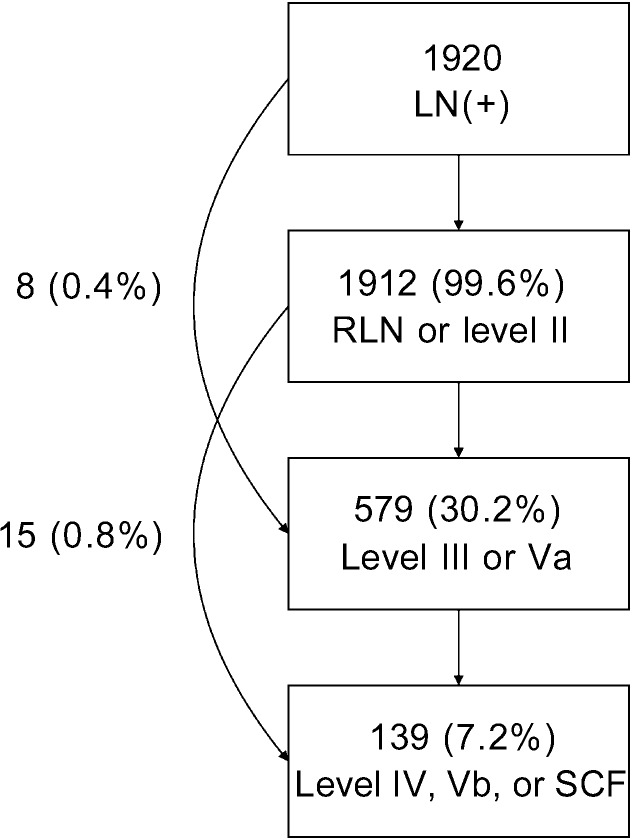
Clinical target volume (CTV) delineation is crucial for tumor control and normal tissue protection. This study aimed to define the locoregional extension patterns of nasopharyngeal carcinoma (NPC) and to improve CTV delineation. Magnetic resonance imaging scans of 2366 newly diagnosed NPC patients were reviewed. According to incidence rates of tumor invasion, the anatomic sites surrounding the nasopharynx were classified into high-risk (>30%), medium-risk (5%-30%), and low-risk (<5%) groups. The lymph node (LN) level was determined according to the Radiation Therapy Oncology Group guidelines, which were further categorized into the upper neck (retropharyngeal region and level II), middle neck (levels III and Va), and lower neck (levels IV and Vb and the supraclavicular fossa). The high-risk anatomic sites were adjacent to the nasopharynx, whereas those at medium-or low-risk were separated from the nasopharynx. If the high-risk anatomic sites were involved, the rates of tumor invasion into the adjacent medium-risk sites increased; if not, the rates were significantly lower (P<0.01). Among the 1920 (81.1%) patients with positive LN, the incidence rates of LN metastasis in the upper, middle, and lower neck were 99.6%, 30.2%, and 7.2%, respectively, and skip metastasis happened in only 1.2% of patients. In the 929 patients who had unilateral upper neck involvement, the rates of contralateral middle neck and lower neck involvement were 1.8% and 0.4%, respectively. Thus, local disease spreads stepwise from proximal sites to distal sites, and LN metastasis spreads from the upper neck to the lower neck. Individualized CTV delineation for NPC may be feasible.
临床靶区(CTV)勾画对于肿瘤控制和正常组织保护至关重要。本研究旨在明确鼻咽癌(NPC)的局部区域扩展模式并改进CTV勾画。回顾了2366例新诊断NPC患者的磁共振成像扫描结果。根据肿瘤侵犯发生率,将鼻咽周围的解剖部位分为高风险(>30%)、中风险(5%-30%)和低风险(<5%)组。淋巴结(LN)水平根据放射治疗肿瘤学组指南确定,进一步分为上颈部(咽后区和Ⅱ区)、中颈部(Ⅲ区和Va区)和下颈部(Ⅳ区和Vb区以及锁骨上窝)。高风险解剖部位紧邻鼻咽,而中风险或低风险部位与鼻咽相隔。如果高风险解剖部位受累,肿瘤侵犯相邻中风险部位的发生率增加;如果未受累,发生率则显著降低(P<0.01)。在1920例(81.1%)LN阳性患者中,上、中、下颈部LN转移的发生率分别为99.6%、30.2%和7.2%,仅1.2%的患者发生跳跃转移。在929例单侧上颈部受累的患者中,对侧中颈部和下颈部受累的发生率分别为1.8%和0.4%。因此,局部病变从近端部位向远端部位逐步扩散,LN转移从上颈部向下颈部扩散。NPC的个体化CTV勾画可能是可行的。