• 文献检索
  • 文档翻译
  • 深度研究
  • 学术资讯
  • Suppr Zotero 插件Zotero 插件
  • 邀请有礼
  • 套餐&价格
  • 历史记录
应用&插件
Suppr Zotero 插件Zotero 插件浏览器插件Mac 客户端Windows 客户端微信小程序
定价
高级版会员购买积分包购买API积分包
服务
文献检索文档翻译深度研究API 文档MCP 服务
关于我们
关于 Suppr公司介绍联系我们用户协议隐私条款
关注我们

Suppr 超能文献

核心技术专利:CN118964589B侵权必究
粤ICP备2023148730 号-1Suppr @ 2026

文献检索

告别复杂PubMed语法,用中文像聊天一样搜索,搜遍4000万医学文献。AI智能推荐,让科研检索更轻松。

立即免费搜索

文件翻译

保留排版,准确专业,支持PDF/Word/PPT等文件格式,支持 12+语言互译。

免费翻译文档

深度研究

AI帮你快速写综述,25分钟生成高质量综述,智能提取关键信息,辅助科研写作。

立即免费体验

鼻咽癌治疗前和治疗后的影像学检查:影像学进展、要点与陷阱

Pre-treatment and post-treatment nasopharyngeal carcinoma imaging: imaging updates, pearls and pitfalls.

作者信息

Li Kwok Yan, Kwok Hoi Ming, Pan Nin Yuan, Cheng Lik Fai, Ma Ka Fai Johnny

机构信息

Department of Diagnostic and Interventional Radiology, Princess Margaret Hospital, Hong Kong SAR, China.

Department of Imaging and Interventional Radiology, Chinese University of Hong Kong, Hong Kong SAR, China.

出版信息

Neuroradiology. 2025 Apr;67(4):1023-1047. doi: 10.1007/s00234-025-03596-z. Epub 2025 Apr 11.

DOI:10.1007/s00234-025-03596-z
PMID:40214770
原文链接:https://pmc.ncbi.nlm.nih.gov/articles/PMC12041163/
Abstract

PURPOSE

Nasopharyngeal carcinoma (NPC) is endemic in Southeast Asia, requiring precise imaging for personalized treatment. This review highlights key imaging challenges and updates from recent literature, emphasizing findings that impact oncological management.

METHODS

We discuss common and uncommon clinical entities, detailing salient imaging features and diagnostic distinctions to aid accurate interpretation.

RESULTS

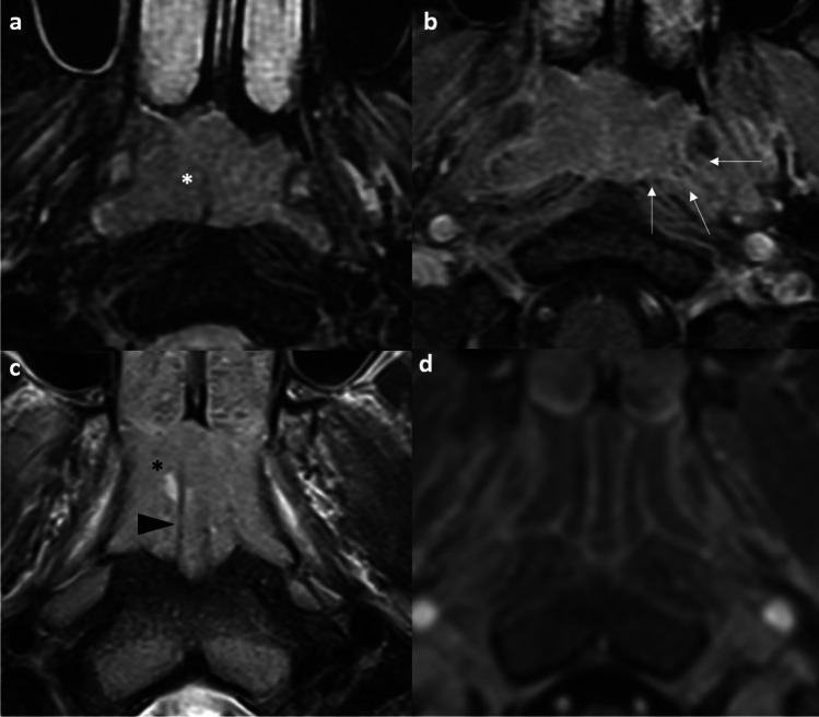
In the pre-treatment setting, leveraging the characteristic MR signals and spread patterns of NPC aids in defining the tumor volume for accurate staging and radiotherapy contouring. Key diagnostic challenges include differentiating tumor from benign hyperplasia, skull base osteomyelitis, and other skull base tumors. Perineural tumor spread, radiological extranodal extension and nodal necrosis further refine primary tumor and nodal assessment. In the post-treatment setting, the key question is whether tumor recurrence exists. Diagnostic challenges involve distinguishing tumor recurrence from scar tissue, post-radiation nasopharyngeal necrosis, or hypertrophied cervical ganglia. For recurrences, endoscopic nasopharyngectomy has emerged as the preferred approach over open surgery or re-irradiation. The text highlights characteristic post-treatment appearances and emphasizes recognizing these patterns to avoid misinterpretation and guide appropriate management.

CONCLUSION

Imaging plays a pivotal role in NPC precision oncology. Mastering imaging pearls and pitfalls empowers radiologists to provide clinicians with reliable, actionable guidance.

摘要

目的

鼻咽癌(NPC)在东南亚地区呈地方性流行,需要精确成像以进行个性化治疗。本综述重点介绍了关键的成像挑战以及近期文献中的更新内容,强调了对肿瘤治疗管理有影响的研究结果。

方法

我们讨论了常见和不常见的临床实体,详细阐述了显著的成像特征和诊断差异,以帮助进行准确解读。

结果

在治疗前阶段,利用鼻咽癌的特征性磁共振信号和扩散模式有助于确定肿瘤体积,以进行准确分期和放疗轮廓勾画。关键的诊断挑战包括区分肿瘤与良性增生、颅底骨髓炎及其他颅底肿瘤。神经周围肿瘤扩散、影像学上的结外扩展和淋巴结坏死进一步完善了原发肿瘤和淋巴结评估。在治疗后阶段,关键问题是是否存在肿瘤复发。诊断挑战包括区分肿瘤复发与瘢痕组织、放疗后鼻咽坏死或颈神经节肥大。对于复发,内镜下鼻咽切除术已成为比开放手术或再次放疗更可取的方法。本文突出了治疗后的特征性表现,并强调识别这些模式以避免误诊并指导适当的治疗管理。

结论

成像在鼻咽癌精准肿瘤学中起着关键作用。掌握成像要点和陷阱使放射科医生能够为临床医生提供可靠且可行的指导。

https://cdn.ncbi.nlm.nih.gov/pmc/blobs/7ee8/12041163/41c5b18cce74/234_2025_3596_Fig17_HTML.jpg
https://cdn.ncbi.nlm.nih.gov/pmc/blobs/7ee8/12041163/0786fffa5d44/234_2025_3596_Fig1_HTML.jpg
https://cdn.ncbi.nlm.nih.gov/pmc/blobs/7ee8/12041163/90ef50f079ed/234_2025_3596_Fig2_HTML.jpg
https://cdn.ncbi.nlm.nih.gov/pmc/blobs/7ee8/12041163/8c177e5e3663/234_2025_3596_Fig3_HTML.jpg
https://cdn.ncbi.nlm.nih.gov/pmc/blobs/7ee8/12041163/f9317918cddc/234_2025_3596_Fig4_HTML.jpg
https://cdn.ncbi.nlm.nih.gov/pmc/blobs/7ee8/12041163/95c3adf16480/234_2025_3596_Fig5_HTML.jpg
https://cdn.ncbi.nlm.nih.gov/pmc/blobs/7ee8/12041163/0f901c74c53a/234_2025_3596_Fig6_HTML.jpg
https://cdn.ncbi.nlm.nih.gov/pmc/blobs/7ee8/12041163/874cab8b0134/234_2025_3596_Fig7_HTML.jpg
https://cdn.ncbi.nlm.nih.gov/pmc/blobs/7ee8/12041163/98d224bf7ea3/234_2025_3596_Fig8_HTML.jpg
https://cdn.ncbi.nlm.nih.gov/pmc/blobs/7ee8/12041163/aa19a8c72900/234_2025_3596_Fig9_HTML.jpg
https://cdn.ncbi.nlm.nih.gov/pmc/blobs/7ee8/12041163/3fb9cbf0d50c/234_2025_3596_Fig10_HTML.jpg
https://cdn.ncbi.nlm.nih.gov/pmc/blobs/7ee8/12041163/4adcb32f7e43/234_2025_3596_Fig11_HTML.jpg
https://cdn.ncbi.nlm.nih.gov/pmc/blobs/7ee8/12041163/77c9622f887a/234_2025_3596_Fig12_HTML.jpg
https://cdn.ncbi.nlm.nih.gov/pmc/blobs/7ee8/12041163/ce869372c3a0/234_2025_3596_Fig13_HTML.jpg
https://cdn.ncbi.nlm.nih.gov/pmc/blobs/7ee8/12041163/d97d55acd150/234_2025_3596_Fig14_HTML.jpg
https://cdn.ncbi.nlm.nih.gov/pmc/blobs/7ee8/12041163/6759eab406bb/234_2025_3596_Fig15_HTML.jpg
https://cdn.ncbi.nlm.nih.gov/pmc/blobs/7ee8/12041163/1670de49392a/234_2025_3596_Fig16_HTML.jpg
https://cdn.ncbi.nlm.nih.gov/pmc/blobs/7ee8/12041163/41c5b18cce74/234_2025_3596_Fig17_HTML.jpg

相似文献

1
Pre-treatment and post-treatment nasopharyngeal carcinoma imaging: imaging updates, pearls and pitfalls.鼻咽癌治疗前和治疗后的影像学检查:影像学进展、要点与陷阱
Neuroradiology. 2025 Apr;67(4):1023-1047. doi: 10.1007/s00234-025-03596-z. Epub 2025 Apr 11.
2
Image-based diagnosis of residual or recurrent nasopharyngeal carcinoma may be a phantom tumor phenomenon.基于影像的残留或复发性鼻咽癌诊断可能是一种假瘤现象。
Medicine (Baltimore). 2021 Feb 26;100(8):e24555. doi: 10.1097/MD.0000000000024555.
3
Management of locally recurrent nasopharyngeal carcinoma.局部复发性鼻咽癌的治疗管理。
Cancer Treat Rev. 2019 Sep;79:101890. doi: 10.1016/j.ctrv.2019.101890. Epub 2019 Aug 21.
4
PET/CT: Nasopharyngeal Cancers.正电子发射断层显像/计算机断层扫描:鼻咽癌
PET Clin. 2022 Apr;17(2):285-296. doi: 10.1016/j.cpet.2021.12.006. Epub 2022 Mar 4.
5
Machine Learning Methods for Optimal Radiomics-Based Differentiation Between Recurrence and Inflammation: Application to Nasopharyngeal Carcinoma Post-therapy PET/CT Images.基于机器学习的最佳放射组学方法区分复发与炎症:在鼻咽癌治疗后 PET/CT 图像中的应用。
Mol Imaging Biol. 2020 Jun;22(3):730-738. doi: 10.1007/s11307-019-01411-9.
6
Skull Base Aspergillus Infection Mimicking Tumor Recurrence: A Nasopharyngeal Carcinoma Case Study.颅底曲霉菌感染模拟肿瘤复发:鼻咽癌病例研究。
Clin Nucl Med. 2024 Aug 1;49(8):750-753. doi: 10.1097/RLU.0000000000005317. Epub 2024 May 26.
7
Pitfalls in the staging of cancer of nasopharyngeal carcinoma.鼻咽癌分期中的陷阱。
Neuroimaging Clin N Am. 2013 Feb;23(1):9-25. doi: 10.1016/j.nic.2012.08.006.
8
Diagnostic value of diffusion-weighted magnetic resonance imaging for local and skull base recurrence of nasopharyngeal carcinoma after radiotherapy.磁共振扩散加权成像对鼻咽癌放疗后局部及颅底复发的诊断价值
Medicine (Baltimore). 2018 Aug;97(34):e11929. doi: 10.1097/MD.0000000000011929.
9
Pretreatment MRI radiomics analysis allows for reliable prediction of local recurrence in non-metastatic T4 nasopharyngeal carcinoma.预处理 MRI 放射组学分析可可靠预测无远处转移 T4 期鼻咽癌的局部复发。
EBioMedicine. 2019 Apr;42:270-280. doi: 10.1016/j.ebiom.2019.03.050. Epub 2019 Mar 27.
10
Intravoxel incoherent motion MRI in differentiation between recurrent carcinoma and postchemoradiation fibrosis of the skull base in patients with nasopharyngeal carcinoma.体素内不相干运动磁共振成像在鼻咽癌患者颅底复发性癌与放化疗后纤维化鉴别中的应用
J Magn Reson Imaging. 2016 Dec;44(6):1556-1564. doi: 10.1002/jmri.25302. Epub 2016 May 26.

本文引用的文献

1
Nasopharyngeal masses in adults-A retrospective analysis of 255 patients to evaluate symptoms, clinical findings, and histological results.成人鼻咽部肿物——对255例患者进行回顾性分析以评估症状、临床检查结果及组织学结果
World J Otorhinolaryngol Head Neck Surg. 2023 Oct 30;11(1):45-51. doi: 10.1002/wjo2.139. eCollection 2025 Mar.
2
Ninth Version of the AJCC and UICC Nasopharyngeal Cancer TNM Staging Classification.美国癌症联合委员会(AJCC)和国际抗癌联盟(UICC)鼻咽癌TNM分期分类第九版。
JAMA Oncol. 2024 Oct 10;10(12):1627-35. doi: 10.1001/jamaoncol.2024.4354.
3
Demystifying the challenging diagnosis of post-radiation nasopharyngeal necrosis on multimodality imaging.
解析多模态影像学检查后放射性鼻咽坏死的诊断难题。
J Med Imaging Radiat Oncol. 2024 Oct;68(7):805-807. doi: 10.1111/1754-9485.13784. Epub 2024 Sep 24.
4
Criteria for the diagnosis of extranodal extension detected on radiological imaging in head and neck cancer: Head and Neck Cancer International Group consensus recommendations.头颈部癌症中影像学检查发现的结外侵犯的诊断标准:头颈部癌症国际协作组共识推荐。
Lancet Oncol. 2024 Jul;25(7):e297-e307. doi: 10.1016/S1470-2045(24)00066-4.
5
Magnetic Resonance Imaging after Nasopharyngeal Endoscopic Resection and Skull Base Reconstruction.鼻咽内镜切除及颅底重建术后的磁共振成像
J Clin Med. 2024 Apr 29;13(9):2624. doi: 10.3390/jcm13092624.
6
Imaging spectrum and complications of otogenic infections: insights into delayed diagnosis.耳源性感染的影像学表现及并发症:延误诊断的相关因素。
Br J Radiol. 2024 Mar 28;97(1156):726-733. doi: 10.1093/bjr/tqae015.
7
Early detection of nasopharyngeal carcinoma: performance of a short contrast-free screening magnetic resonance imaging.早期鼻咽癌检测:短无对比增强筛查磁共振成像的性能。
J Natl Cancer Inst. 2024 May 8;116(5):665-672. doi: 10.1093/jnci/djad260.
8
Radiologic extranodal extension for nodal staging in nasopharyngeal carcinoma.鼻咽癌淋巴结分期的影像学结外延伸。
Radiother Oncol. 2024 Feb;191:110050. doi: 10.1016/j.radonc.2023.110050. Epub 2023 Dec 13.
9
Differentiating nasopharyngeal carcinoma from lymphoma in the head and neck region using the apparent diffusion coefficient (ADC) value: a systematic review and meta-analysis.利用表观扩散系数(ADC)值鉴别头颈部区域的鼻咽癌与淋巴瘤:一项系统评价和荟萃分析。
Pol J Radiol. 2023 Oct 17;88:e472-e482. doi: 10.5114/pjr.2023.132172. eCollection 2023.
10
Nasopharyngeal Cancer Incidence and Mortality in 185 Countries in 2020 and the Projected Burden in 2040: Population-Based Global Epidemiological Profiling.2020 年 185 个国家和地区的鼻咽癌发病和死亡情况以及 2040 年的预测负担:基于人群的全球流行病学特征描述。
JMIR Public Health Surveill. 2023 Sep 20;9:e49968. doi: 10.2196/49968.