Division of Radiation Oncology, University Hospital, Geneva, Switzerland.
Radiat Oncol. 2012 Aug 8;7:134. doi: 10.1186/1748-717X-7-134.
To assess the influence of sentinel lymph nodes (SNs) SPECT/CT and 18 F-choline (18 F-FCH) PET/CT in radiotherapy (RT) treatment planning for prostate cancer patients with a high-risk for lymph node (LN) involvement.
Twenty high-risk prostate cancer patients underwent a pelvic SPECT acquisition following a transrectal ultrasound guided injection of 99mTc-Nanocoll into the prostate. In all patients but one an 18 F-FCH PET/CT for RT treatment planning was performed. SPECT studies were coregistered with the respective abdominal CTs. Pelvic SNs localized on SPECT/CT and LN metastases detected by 18 F-FCH PET/CT were compared to standard pelvic clinical target volumes (CTV).
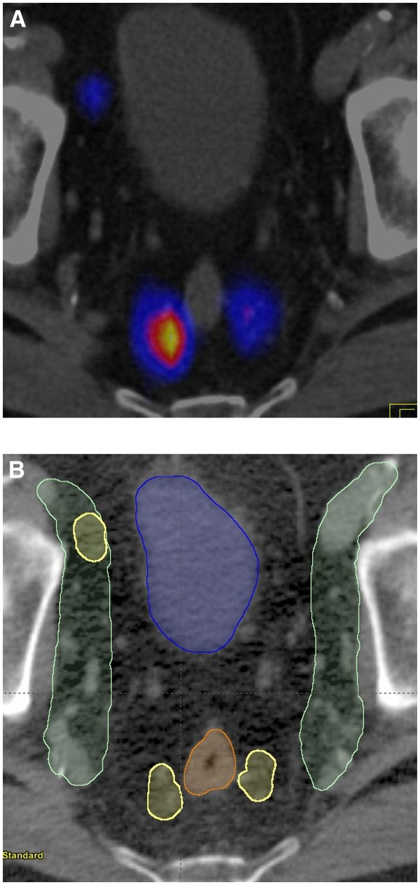
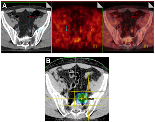
A total of 104 pelvic SNs were identified on SPECT/CT (mean 5.2 SNs/patient; range 1-10). Twenty-seven SNs were located outside the standard pelvic CTV, 17 in the proximal common iliac and retroperitoneal regions above S1, 9 in the pararectal fat and 1 in the inguinal region. SPECT/CT succeeded to optimize the definition of the CTV and treatment plans in 6/20 patients due to the presence of pararectal SNs located outside the standard treatment volume. 18 F-FCH PET/CT identified abnormal tracer uptake in the iliac LN region in 2/19 patients. These abnormal LNs were negative on SPECT/CT suggesting a potential blockade of lymphatic drainage by metastatic LNs with a high tumour burden.
Multimodality imaging which combines SPECT/CT prostate lymphoscintigraphy and 18 F-FCH PET/CT identified SNs outside standard pelvic CTVs or highly suspicious pelvic LNs in 40% of high-risk prostate cancer patients, highlighting the potential impact of this approach in RT treatment planning.
评估前哨淋巴结 (SN) SPECT/CT 和 18 F-胆碱 (18 F-FCH) PET/CT 对高危淋巴结 (LN) 受累前列腺癌患者放射治疗 (RT) 治疗计划的影响。
20 例高危前列腺癌患者在经直肠超声引导下将 99mTc-Nanocoll 注射入前列腺后进行盆腔 SPECT 采集。除 1 例患者外,所有患者均进行 18 F-FCH PET/CT 进行 RT 治疗计划。SPECT 研究与各自的腹部 CT 进行配准。在 SPECT/CT 上定位的盆腔 SN 和 18 F-FCH PET/CT 检测到的 LN 转移与标准盆腔临床靶区 (CTV) 进行比较。
SPECT/CT 共识别出 104 个盆腔 SN(平均每个患者 5.2 个 SN;范围 1-10)。27 个 SN 位于标准盆腔 CTV 之外,17 个位于 S1 上方的近端髂总动脉和腹膜后区域,9 个位于直肠旁脂肪中,1 个位于腹股沟区域。由于标准治疗体积外存在直肠旁 SN,SPECT/CT 成功优化了 6/20 例患者的 CTV 和治疗计划。18 F-FCH PET/CT 在 19 例患者中的髂 LN 区域发现异常示踪剂摄取。这些异常 LN 在 SPECT/CT 上呈阴性,提示高肿瘤负荷的转移性 LN 可能阻断了淋巴引流。
结合 SPECT/CT 前列腺淋巴闪烁显像和 18 F-FCH PET/CT 的多模态成像,在 40%的高危前列腺癌患者中识别出标准盆腔 CTV 外的 SN 或高度可疑的盆腔 LN,突出了这种方法在 RT 治疗计划中的潜在影响。