• 文献检索
  • 文档翻译
  • 深度研究
  • 学术资讯
  • Suppr Zotero 插件Zotero 插件
  • 邀请有礼
  • 套餐&价格
  • 历史记录
应用&插件
Suppr Zotero 插件Zotero 插件浏览器插件Mac 客户端Windows 客户端微信小程序
定价
高级版会员购买积分包购买API积分包
服务
文献检索文档翻译深度研究API 文档MCP 服务
关于我们
关于 Suppr公司介绍联系我们用户协议隐私条款
关注我们

Suppr 超能文献

核心技术专利:CN118964589B侵权必究
粤ICP备2023148730 号-1Suppr @ 2026

文献检索

告别复杂PubMed语法,用中文像聊天一样搜索,搜遍4000万医学文献。AI智能推荐,让科研检索更轻松。

立即免费搜索

文件翻译

保留排版,准确专业,支持PDF/Word/PPT等文件格式,支持 12+语言互译。

免费翻译文档

深度研究

AI帮你快速写综述,25分钟生成高质量综述,智能提取关键信息,辅助科研写作。

立即免费体验

前列腺淋巴结分布:一份源自PET/CT的前列腺癌患者手术治疗前后的解剖图谱。

Distribution of prostate nodes: a PET/CT-derived anatomic atlas of prostate cancer patients before and after surgical treatment.

作者信息

Hegemann Nina-Sophie, Wenter Vera, Spath Sonja, Kusumo Nadia, Li Minglun, Bartenstein Peter, Fendler Wolfgang P, Stief Christian, Belka Claus, Ganswindt Ute

机构信息

Department of Radiation Oncology, Klinikum der Universität, Ludwig-Maximilians-University (LMU), Marchioninistrasse 15, 81377, Munich, Germany.

Department of Nuclear Medicine, Klinikum der Universität, Ludwig-Maximilians-University (LMU), Marchioninistrasse 15, 81377, Munich, Germany.

出版信息

Radiat Oncol. 2016 Mar 11;11:37. doi: 10.1186/s13014-016-0615-9.

DOI:10.1186/s13014-016-0615-9
PMID:26968955
原文链接:https://pmc.ncbi.nlm.nih.gov/articles/PMC4788881/
Abstract

BACKGROUND

In order to define adequate radiation portals in nodal positive prostate cancer a detailed knowledge of the anatomic lymph-node distribution is mandatory. We therefore systematically analyzed the localization of Choline PET/CT positive lymph nodes and compared it to the RTOG recommendation of pelvic CTV, as well as to previous work, the SPECT sentinel lymph node atlas.

METHODS

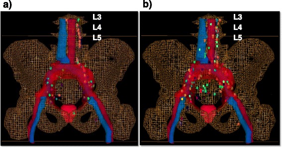
Thirty-two patients being mostly high risk patients with a PSA of 12.5 ng/ml (median) received PET/CT before any treatment. Eighty-seven patients received PET/CT for staging due to biochemical failure with a median PSA of 3.12 ng/ml. Each single PET-positive lymph node was manually contoured in a "virtual" patient dataset to achieve a 3-D visualization, resulting in an atlas of the cumulative PET positive lymph node distribution. Further the PET-positive lymph node location in each patient was assessed with regard to the existence of a potential geographic miss (i.e. PET-positive lymph nodes that would not have been treated adequately by the RTOG consensus on CTV definition of pelvic lymph nodes).

RESULTS

Seventy-eight and 209 PET positive lymph nodes were detected in patients with no prior treatment and in postoperative patients, respectively. The most common sites of PET positive lymph nodes in patients with no prior treatment were external iliac (32.1 %), followed by common iliac (23.1 %) and para-aortic (19.2 %). In postoperative patients the most common sites of PET positive lymph nodes were common iliac (24.9 %), followed by external iliac (23.0 %) and para-aortic (20.1 %). In patients with no prior treatment there were 34 (43.6 %) and in postoperative patients there were 77 (36.8 %) of all detected lymph nodes that would not have been treated adequately using the RTOG CTV. We compared the distribution of lymph nodes gained by Choline PET/CT to the preexisting SPECT sentinel lymph node atlas and saw an overall good congruence.

CONCLUSIONS

Choline PET/CT and SPECT sentinel lymph node atlas are comparable to each other. More than one-third of the PET positive lymph nodes in patients with no prior treatment and in postoperative patients would not have been treated adequately using the RTOG CTV. To reduce geographical miss, image based definition of an individual target volume is necessary.

摘要

背景

为了确定淋巴结阳性前列腺癌的合适放疗野,必须详细了解解剖学上的淋巴结分布情况。因此,我们系统地分析了胆碱正电子发射断层扫描/计算机断层扫描(Choline PET/CT)阳性淋巴结的定位,并将其与美国放射肿瘤学会(RTOG)关于盆腔临床靶体积(CTV)的建议以及先前的研究——SPECT前哨淋巴结图谱进行比较。

方法

32例大多为高危患者,中位前列腺特异性抗原(PSA)为12.5 ng/ml,在接受任何治疗前接受了PET/CT检查。87例因生化复发接受PET/CT分期检查的患者,中位PSA为3.12 ng/ml。在一个“虚拟”患者数据集中,对每个PET阳性淋巴结进行手动勾勒轮廓,以实现三维可视化,从而得到累积PET阳性淋巴结分布图谱。此外,针对每位患者的PET阳性淋巴结位置,评估是否存在潜在的区域遗漏(即根据RTOG关于盆腔淋巴结CTV定义的共识,未得到充分治疗的PET阳性淋巴结)。

结果

在未接受过治疗的患者和术后患者中,分别检测到78个和209个PET阳性淋巴结。未接受过治疗的患者中,PET阳性淋巴结最常见的部位是髂外淋巴结(32.1%),其次是髂总淋巴结(23.1%)和腹主动脉旁淋巴结(19.2%)。术后患者中,PET阳性淋巴结最常见的部位是髂总淋巴结(24.9%),其次是髂外淋巴结(23.0%)和腹主动脉旁淋巴结(20.1%)。在未接受过治疗的患者中,使用RTOG CTV无法得到充分治疗的所有检测到的淋巴结有34个(43.6%);在术后患者中,这一比例为77个(36.8%)。我们将胆碱PET/CT获得的淋巴结分布与现有的SPECT前哨淋巴结图谱进行比较,发现总体一致性良好。

结论

胆碱PET/CT和SPECT前哨淋巴结图谱具有可比性。未接受过治疗的患者和术后患者中,超过三分之一的PET阳性淋巴结使用RTOG CTV无法得到充分治疗。为了减少区域遗漏,有必要基于图像定义个体靶体积。

https://cdn.ncbi.nlm.nih.gov/pmc/blobs/2af6/4788881/96e9f005fd46/13014_2016_615_Fig2_HTML.jpg
https://cdn.ncbi.nlm.nih.gov/pmc/blobs/2af6/4788881/557a6d7cc793/13014_2016_615_Fig1_HTML.jpg
https://cdn.ncbi.nlm.nih.gov/pmc/blobs/2af6/4788881/96e9f005fd46/13014_2016_615_Fig2_HTML.jpg
https://cdn.ncbi.nlm.nih.gov/pmc/blobs/2af6/4788881/557a6d7cc793/13014_2016_615_Fig1_HTML.jpg
https://cdn.ncbi.nlm.nih.gov/pmc/blobs/2af6/4788881/96e9f005fd46/13014_2016_615_Fig2_HTML.jpg

相似文献

1
Distribution of prostate nodes: a PET/CT-derived anatomic atlas of prostate cancer patients before and after surgical treatment.前列腺淋巴结分布:一份源自PET/CT的前列腺癌患者手术治疗前后的解剖图谱。
Radiat Oncol. 2016 Mar 11;11:37. doi: 10.1186/s13014-016-0615-9.
2
High occurrence of aberrant lymph node spread on magnetic resonance lymphography in prostate cancer patients with a biochemical recurrence after radical prostatectomy.根治性前列腺切除术后生化复发的前列腺癌患者,磁共振淋巴造影显示异常淋巴结转移发生率高。
Int J Radiat Oncol Biol Phys. 2012 Mar 15;82(4):1405-10. doi: 10.1016/j.ijrobp.2011.04.054. Epub 2011 Jun 2.
3
Histological verification of 11C-choline-positron emission/computed tomography-positive lymph nodes in patients with biochemical failure after treatment for localized prostate cancer.局限性前列腺癌治疗后生化复发患者中11C-胆碱正电子发射/计算机断层扫描阳性淋巴结的组织学验证
BJU Int. 2008 Aug;102(4):446-51. doi: 10.1111/j.1464-410X.2008.07592.x. Epub 2008 Apr 11.
4
Patterns of lymph node positivity on C-acetate PET imaging in correlation to the RTOG pelvic radiation field for prostate cancer.C-乙酰酸盐 PET 成像中与前列腺癌 RTOG 盆部放疗野相关的淋巴结阳性模式。
Pract Radiat Oncol. 2017 Sep-Oct;7(5):325-331. doi: 10.1016/j.prro.2017.03.002. Epub 2017 Mar 6.
5
Targeted salvage lymphadenectomy in patients treated with radical prostatectomy with biochemical recurrence: complete biochemical response without adjuvant therapy in patients with low volume lymph node recurrence over a long-term follow-up.接受根治性前列腺切除术后出现生化复发的患者的靶向挽救性淋巴结清扫术:长期随访中,低容量淋巴结复发患者在无辅助治疗情况下实现完全生化缓解。
BMC Urol. 2015 Feb 21;15(1):10. doi: 10.1186/s12894-015-0004-y.
6
Pattern of occult nodal relapse diagnosed with (18)F-fluoro-choline PET/CT in prostate cancer patients with biochemical failure after prostate-only radiotherapy.仅接受前列腺放疗后出现生化失败的前列腺癌患者中,通过(18)F-氟胆碱PET/CT诊断出的隐匿性淋巴结复发模式。
Radiother Oncol. 2014 Apr;111(1):120-5. doi: 10.1016/j.radonc.2014.03.008. Epub 2014 Apr 26.
7
Target volume definition in high-risk prostate cancer patients using sentinel node SPECT/CT and 18 F-choline PET/CT.高危前列腺癌患者应用前哨淋巴结 SPECT/CT 和 18 F-胆碱 PET/CT 进行靶区定义。
Radiat Oncol. 2012 Aug 8;7:134. doi: 10.1186/1748-717X-7-134.
8
Detection of lymph-node metastases with integrated [11C]choline PET/CT in patients with PSA failure after radical retropubic prostatectomy: results confirmed by open pelvic-retroperitoneal lymphadenectomy.耻骨后根治性前列腺切除术后前列腺特异性抗原(PSA)失败患者中,采用[11C]胆碱PET/CT融合成像检测淋巴结转移:开放盆腔-腹膜后淋巴结清扫术证实结果
Eur Urol. 2007 Aug;52(2):423-9. doi: 10.1016/j.eururo.2007.03.032. Epub 2007 Mar 20.
9
Does Choline PET/CT Change the Management of Prostate Cancer Patients With Biochemical Failure?胆碱正电子发射断层扫描/计算机断层扫描(Choline PET/CT)是否会改变生化复发的前列腺癌患者的治疗管理?
Am J Clin Oncol. 2017 Jun;40(3):256-259. doi: 10.1097/COC.0000000000000139.
10
Influence of (11)C-choline PET/CT on the treatment planning for salvage radiation therapy in patients with biochemical recurrence of prostate cancer.(11)C-胆碱 PET/CT 对前列腺癌生化复发患者挽救性放疗计划制定的影响。
Radiother Oncol. 2011 May;99(2):193-200. doi: 10.1016/j.radonc.2011.05.005. Epub 2011 May 26.

引用本文的文献

1
Necessity of Pelvic Lymph Node Irradiation in Patients with Recurrent Prostate Cancer after Radical Prostatectomy in the PSMA PET/CT Era: A Narrative Review.PSMA PET/CT时代前列腺癌根治术后复发性前列腺癌患者盆腔淋巴结照射的必要性:一项叙述性综述
Biomedicines. 2022 Dec 24;11(1):38. doi: 10.3390/biomedicines11010038.
2
Patterns of Lymph Node Failure in Patients With Recurrent Prostate Cancer Postradical Prostatectomy and Implications for Salvage Therapies.前列腺癌根治术后复发患者的淋巴结转移模式及其对挽救性治疗的意义
Adv Radiat Oncol. 2020 Aug 31;5(6):1126-1140. doi: 10.1016/j.adro.2020.07.009. eCollection 2020 Nov-Dec.
3

本文引用的文献

1
Prostate cancer: Identifying sites of recurrence with choline-PET-CT imaging.前列腺癌:利用胆碱正电子发射断层扫描-计算机断层扫描成像识别复发部位。
Nat Rev Urol. 2015 Mar;12(3):134-5. doi: 10.1038/nrurol.2014.321. Epub 2014 Dec 2.
2
The diagnostic value of PET/CT imaging with the (68)Ga-labelled PSMA ligand HBED-CC in the diagnosis of recurrent prostate cancer.(68)镓标记的前列腺特异性膜抗原(PSMA)配体HBED-CC的PET/CT成像在复发性前列腺癌诊断中的价值
Eur J Nucl Med Mol Imaging. 2015 Feb;42(2):197-209. doi: 10.1007/s00259-014-2949-6. Epub 2014 Nov 20.
3
Individualized image-based lymph node irradiation for prostate cancer.
A CT-based radiomics model to detect prostate cancer lymph node metastases in PSMA radioguided surgery patients.
基于 CT 的放射组学模型可用于探测 PSMA 放射性引导手术患者的前列腺癌淋巴结转移。
Eur J Nucl Med Mol Imaging. 2020 Dec;47(13):2968-2977. doi: 10.1007/s00259-020-04864-1. Epub 2020 May 28.
4
Haute Couture or Ready-to-Wear? Tailored Pelvic Radiotherapy for Prostate Cancer Based on Individualized Sentinel Lymph Node Detection.高级定制还是成衣?基于个体化前哨淋巴结检测的前列腺癌定制盆腔放疗。
Cancers (Basel). 2020 Apr 10;12(4):944. doi: 10.3390/cancers12040944.
5
Good clinical practice recommendations for the use of PET/CT in oncology.关于在肿瘤学中使用PET/CT的良好临床实践建议。
Eur J Nucl Med Mol Imaging. 2020 Jan;47(1):28-50. doi: 10.1007/s00259-019-04553-8. Epub 2019 Oct 21.
6
Ga-PSMA-11 PET/CT Mapping of Prostate Cancer Biochemical Recurrence After Radical Prostatectomy in 270 Patients with a PSA Level of Less Than 1.0 ng/mL: Impact on Salvage Radiotherapy Planning.在 PSA 水平低于 1.0ng/ml 的 270 例患者中,Ga-PSMA-11 PET/CT 对前列腺癌根治术后生化复发的定位:对挽救性放疗计划的影响。
J Nucl Med. 2018 Feb;59(2):230-237. doi: 10.2967/jnumed.117.201749. Epub 2017 Nov 9.
7
Patterns of failure after radical prostatectomy in prostate cancer - implications for radiation therapy planning after Ga-PSMA-PET imaging.前列腺癌根治性前列腺切除术后的失败模式——对镓-PSMA-PET成像后放射治疗计划的影响
Eur J Nucl Med Mol Imaging. 2017 Sep;44(10):1656-1662. doi: 10.1007/s00259-017-3746-9. Epub 2017 Jun 23.
基于个体化图像的前列腺癌淋巴结照射。
Nat Rev Urol. 2013 Jul;10(7):376-85. doi: 10.1038/nrurol.2013.111. Epub 2013 May 28.
4
The role of 11C-choline and 18F-fluorocholine positron emission tomography (PET) and PET/CT in prostate cancer: a systematic review and meta-analysis.11C-胆碱和 18F-氟胆碱正电子发射断层扫描(PET)和 PET/CT 在前列腺癌中的作用:系统评价和荟萃分析。
Eur Urol. 2013 Jul;64(1):106-17. doi: 10.1016/j.eururo.2013.04.019. Epub 2013 Apr 19.
5
Geographical distribution of lymph node metastases on MR lymphography in prostate cancer patients.前列腺癌患者磁共振淋巴造影中淋巴结转移的地理分布。
Radiother Oncol. 2013 Jan;106(1):59-63. doi: 10.1016/j.radonc.2012.10.021. Epub 2012 Nov 27.
6
18F-Fluoroethylcholine PET/CT identifies lymph node metastasis in patients with prostate-specific antigen failure after radical prostatectomy but underestimates its extent.18F-氟乙基胆碱 PET/CT 可识别根治性前列腺切除术后 PSA 失败患者的淋巴结转移,但低估了其范围。
Eur Urol. 2013 May;63(5):792-6. doi: 10.1016/j.eururo.2012.08.003. Epub 2012 Aug 10.
7
Mapping of pelvic lymph node metastases in prostate cancer.前列腺癌盆腔淋巴结转移的定位。
Eur Urol. 2013 Mar;63(3):450-8. doi: 10.1016/j.eururo.2012.06.057. Epub 2012 Jul 6.
8
Distribution of prostate sentinel nodes: a SPECT-derived anatomic atlas.前列腺前哨淋巴结的分布:SPECT 衍生解剖图谱。
Int J Radiat Oncol Biol Phys. 2011 Apr 1;79(5):1364-72. doi: 10.1016/j.ijrobp.2010.01.012. Epub 2010 Aug 25.
9
RTOG GU Radiation oncology specialists reach consensus on pelvic lymph node volumes for high-risk prostate cancer.放射治疗肿瘤学组(RTOG)泌尿生殖系统放射肿瘤学专家就高危前列腺癌的盆腔淋巴结体积达成共识。
Int J Radiat Oncol Biol Phys. 2009 Jun 1;74(2):383-7. doi: 10.1016/j.ijrobp.2008.08.002. Epub 2008 Oct 22.
10
Optimized coverage of high-risk adjuvant lymph node areas in prostate cancer using a sentinel node-based, intensity-modulated radiation therapy technique.使用基于前哨淋巴结的调强放射治疗技术优化前列腺癌高危辅助淋巴结区域的覆盖范围。
Int J Radiat Oncol Biol Phys. 2007 Feb 1;67(2):347-55. doi: 10.1016/j.ijrobp.2006.08.082.