Liu Ting, Zhai Hualei, Xu Yuanyuan, Dong Yanling, Sun Yajie, Zang Xinjie, Zhao Jing
State Key Laboratory Cultivation Base, Shandong Provincial Key Laboratory of Ophthalmology, Shandong Eye Institute, Shandong Academy of Medical Sciences, Qingdao, China.
Mol Vis. 2012;18:2137-46. Epub 2012 Jul 26.
Severe chemical burns can cause necrosis of ocular surface tissues following the infiltration of inflammatory cells. It has been shown that amniotic membrane transplantation (AMT) is an effective treatment for severe chemical burns, but the phenotypes of cells that infiltrate the amniotic membrane and the clinical significance of these cellular infiltrations have not previously been reported. The present work studies the inflammation cell traps and apoptosis inducing roles of the amniotic membrane after AMT in patients with acute chemical burns.
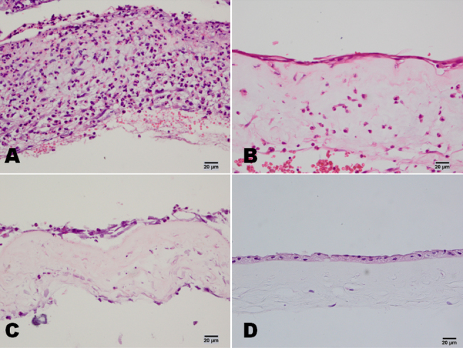
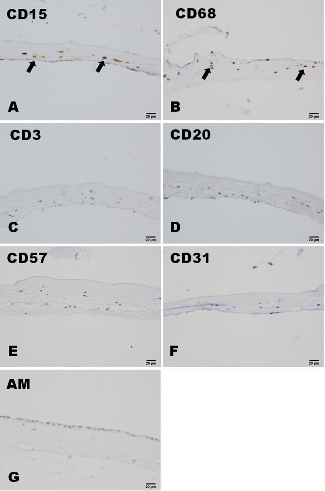
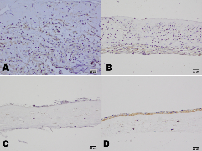
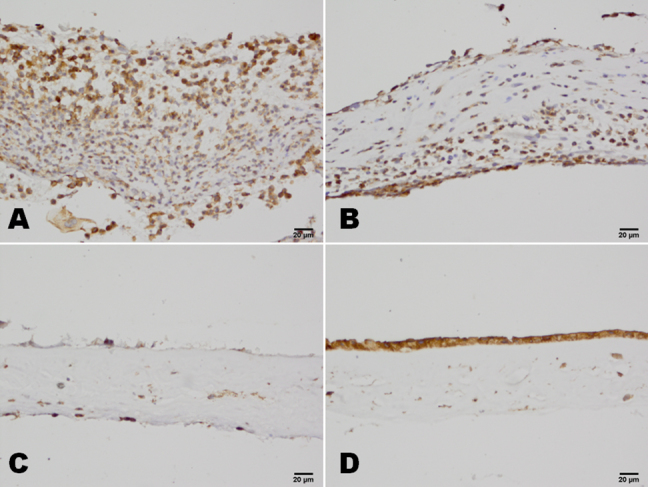
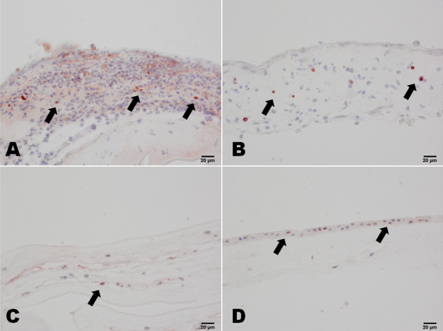
A total of 30 patients with acute alkaline burns were classified as having either moderate or severe burns. In all participants, AMT was performed within one week of his/her injury. After 7-9 days, the transplanted amniotic membranes were removed. Histopathological and immunohistochemical techniques were used for the examination and detection of infiltrating cells, and tests for the expression of CD (cluster of differentiation)15, CD68, CD3, CD20, CD57, CD31, CD147, and CD95 (Fas) were performed. A TUNEL (TdT-mediated dUTP nick end labeling) assay was used to confirm apoptosis of the infiltrating cells. Three patients with herpes simplex-induced keratitis who had undergone AMT to treat persistent epithelium defects were used as a control group. Amniotic membrane before transplantation was used as another control.
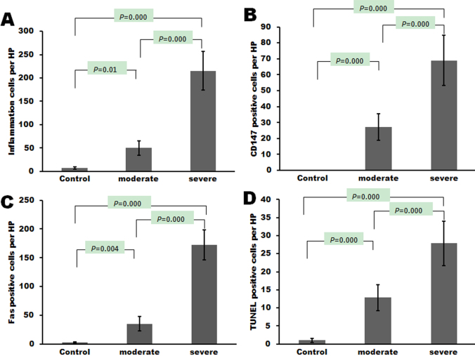
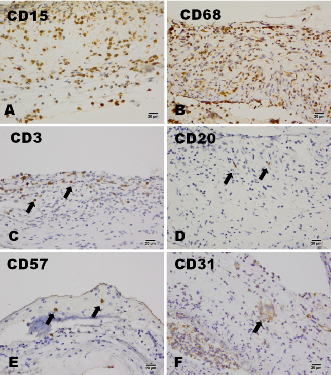
After amniotic membrane transplantation, the number of infiltrating cells in patients with severe burns was significantly higher than in patients with moderate burns or in control patients (p<0.05). Among the severe burns patients, CD15 and CD68 were widely expressed in the infiltrating cells, and CD3, CD20, and CD57 were only found in a small number of cells. Occasionally, CD31-positive cells were found in the amniotic membranes. More cells that were CD147, Fas, and TUNEL positive were found in patients with severe burns than in patients with moderate burns or in control patients.
Neutrophils and macrophages were the main cells that had infiltrated into the amniotic membrane during the acute phase of healing from a chemical burns. AMT can trap different inflammatory cells and induce apoptosis of inflammatory cells in acute ocular chemical burns.
严重化学烧伤可导致炎性细胞浸润后眼表组织坏死。已表明羊膜移植(AMT)是治疗严重化学烧伤的有效方法,但此前尚未报道浸润羊膜的细胞表型以及这些细胞浸润的临床意义。本研究探讨急性化学烧伤患者行AMT后羊膜的炎症细胞捕获及诱导凋亡作用。
30例急性碱性烧伤患者分为中度或重度烧伤。所有参与者均在受伤后1周内进行AMT。7 - 9天后,取出移植的羊膜。采用组织病理学和免疫组织化学技术检查和检测浸润细胞,并检测CD(分化簇)15、CD68、CD3、CD20、CD57、CD31、CD147和CD95(Fas)的表达。采用TUNEL(末端脱氧核苷酸转移酶介导的dUTP缺口末端标记)法确认浸润细胞的凋亡。3例因单纯疱疹性角膜炎行AMT治疗持续性上皮缺损的患者作为对照组。移植前的羊膜作为另一对照组。
羊膜移植后,重度烧伤患者的浸润细胞数量显著高于中度烧伤患者或对照组患者(p<0.05)。在重度烧伤患者中,CD15和CD68在浸润细胞中广泛表达,而CD3、CD20和CD57仅在少数细胞中发现。偶尔在羊膜中发现CD31阳性细胞。重度烧伤患者中CD147、Fas和TUNEL阳性的细胞比中度烧伤患者或对照组患者更多。
中性粒细胞和巨噬细胞是化学烧伤愈合急性期浸润羊膜的主要细胞。AMT可捕获不同的炎性细胞并诱导急性眼化学烧伤中炎性细胞的凋亡。