Department of Pathology, University of Göttingen, Robert-Koch-Str 40, 37083 Göttingen, Germany.
Diagn Pathol. 2012 Aug 13;7:95. doi: 10.1186/1746-1596-7-95.
Papillary renal cell carcinoma (RCC) represents a rare tumor, which is divided, based on histological criteria, into two subtypes. In contrast to type I papillary RCC type II papillary RCC shows a worse prognosis. So far, reliable immunohistochemical markers for the distinction of these subtypes are not available.
In the present study the expression of N(neural)-, E(epithelial)-, P(placental)-, and KSP(kidney specific)-cadherin was examined in 22 papillary RCC of histological type I and 18 papillary RCC of histological type II (n = 40).
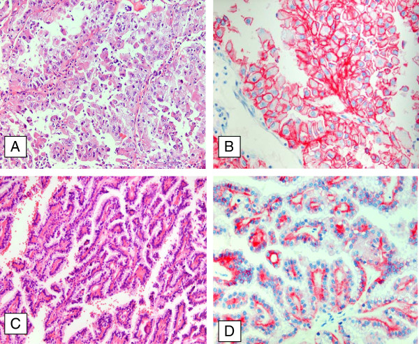
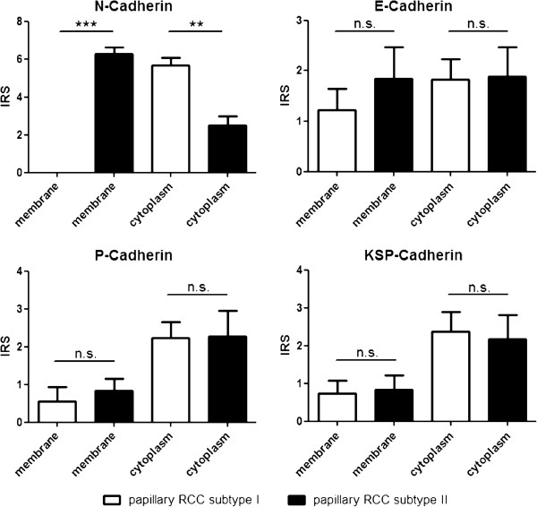
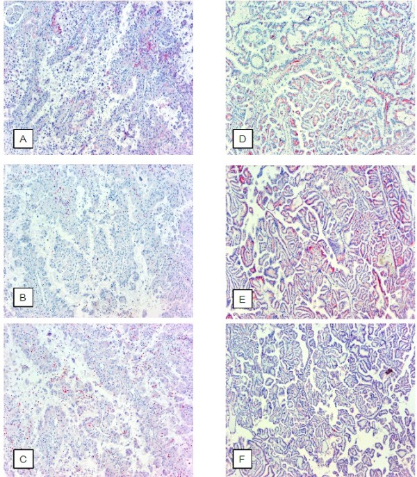
All papillary RCC type II displayed a membranous expression for N-cadherin, whereas type I did not show any membranous positivity for N-cadherin. E-cadherin exhibited a stronger, but not significant, membranous as well as cytoplasmic expression in type II than in type I papillary RCC. A diagnostic relevant expression of P- and KSP-cadherin could not be demonstrated in both tumor entities.
Thus N-cadherin represents the first immunhistochemical marker for a clear cut differentiation between papillary RCC type I and type II and could be a target for therapy and diagnostic in the future.
The virtual slide(s) for this article can be found here: http://www.diagnosticpathology.diagnomx.eu/vs/2011556982761733.
乳头状肾细胞癌(RCC)是一种罕见的肿瘤,根据组织学标准可分为两型。与 I 型乳头状 RCC 相反,II 型乳头状 RCC 预后较差。到目前为止,还没有可靠的免疫组织化学标志物来区分这两种亚型。
本研究检测了 22 例组织学 I 型和 18 例组织学 II 型乳头状 RCC(n=40)中 N(神经)、E(上皮)、P(胎盘)和 KSP(肾脏特异)-钙黏蛋白的表达。
所有 II 型乳头状 RCC 均显示 N-钙黏蛋白的膜表达,而 I 型则无任何膜阳性。与 I 型乳头状 RCC 相比,E-钙黏蛋白在 II 型中表现出更强的膜和细胞质表达,但不显著。在两种肿瘤实体中均未能证明 P-和 KSP-钙黏蛋白有诊断相关的表达。
因此,N-钙黏蛋白是区分 I 型和 II 型乳头状 RCC 的第一个免疫组织化学标志物,可能成为未来治疗和诊断的靶点。
本文的虚拟幻灯片可以在这里找到:http://www.diagnosticpathology.diagnomx.eu/vs/2011556982761733。