Biology Department, Gannon University, Erie, PA 16541, USA.
BMC Microbiol. 2012 Aug 15;12:176. doi: 10.1186/1471-2180-12-176.
Chlamydia trachomatis is an intracellular bacterium that resides in the conjunctival and reproductive tract mucosae and is responsible for an array of acute and chronic diseases. A percentage of these infections persist even after use of antibiotics, suggesting the need for alternative treatments. Previous studies have demonstrated anti-bacterial effects using different wavelengths of visible light at varying energy densities, though only against extracellular bacteria. We investigated the effects of visible light (405 and 670 nm) irradiation via light emitting diode (LEDs) on chlamydial growth in endocervical epithelial cells, HeLa, during active and penicillin-induced persistent infections. Furthermore, we analyzed the effect of this photo treatment on the ensuing secretion of IL-6 and CCL2, two pro-inflammatory cytokines that have previously been identified as immunopathologic components associated with trichiasis in vivo.
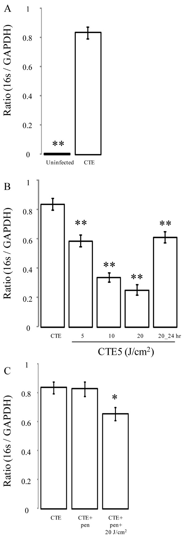
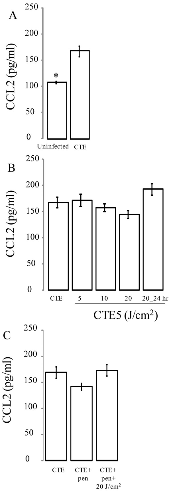
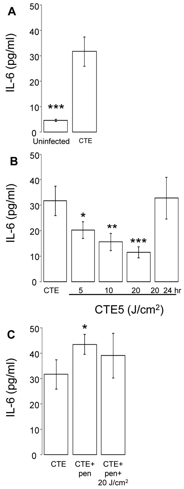
C. trachomatis-infected HeLa cells were treated with 405 or 670 nm irradiation at varying energy densities (0 - 20 J/cm2). Bacterial growth was assessed by quantitative real-time PCR analyzing the 16S: GAPDH ratio, while cell-free supernatants were examined for IL-6 and monocyte chemoattractant protein-1 (CCL2) production. Our results demonstrated a significant dose-dependent inhibitory effect on chlamydial growth during both active and persistent infections following 405 nm irradiation. Diminished bacterial load corresponded to lower IL-6 concentrations, but was not related to CCL2 levels. In vitro modeling of a persistent C. trachomatis infection induced by penicillin demonstrated significantly elevated IL-6 levels compared to C. trachomatis infection alone, though 405 nm irradiation had a minimal effect on this production.
Together these results identify novel inhibitory effects of 405 nm violet light on the bacterial growth of intracellular bacterium C. trachomatis in vitro, which also coincides with diminished levels of the pro-inflammatory cytokine IL-6.
沙眼衣原体是一种细胞内细菌,存在于结膜和生殖道黏膜中,可引起一系列急性和慢性疾病。尽管使用了抗生素,仍有一定比例的感染持续存在,这表明需要替代治疗。先前的研究已经证明,使用不同波长和不同能量密度的可见光可以产生抗菌作用,尽管这些研究只针对细胞外细菌。我们研究了 405nm 和 670nm 可见光(通过发光二极管 LED 照射)对宫颈上皮细胞、HeLa 细胞中沙眼衣原体生长的影响,包括在活性感染和青霉素诱导的持续感染期间。此外,我们分析了这种光疗对随后分泌白细胞介素 6(IL-6)和 C 趋化因子配体 2(CCL2)的影响,这两种促炎细胞因子先前被鉴定为与体内三刺线虫病相关的免疫病理成分。
用 405nm 或 670nm 照射不同能量密度(0-20J/cm2)处理沙眼衣原体感染的 HeLa 细胞。通过定量实时 PCR 分析 16S:GAPDH 比值来评估细菌生长情况,同时检查细胞培养液中白细胞介素 6(IL-6)和单核细胞趋化蛋白 1(CCL2)的分泌情况。结果表明,405nm 照射对活性感染和持续感染期间沙眼衣原体的生长具有显著的剂量依赖性抑制作用。细菌负荷的降低与 IL-6 浓度降低相关,但与 CCL2 水平无关。与单独的沙眼衣原体感染相比,青霉素诱导的持续沙眼衣原体感染的体外模型显示 IL-6 水平显著升高,而 405nm 照射对这种产生的影响很小。
这些结果共同确定了 405nm 紫光对细胞内细菌沙眼衣原体在体外的细菌生长具有新的抑制作用,这也与促炎细胞因子 IL-6 的水平降低有关。