• 文献检索
  • 文档翻译
  • 深度研究
  • 学术资讯
  • Suppr Zotero 插件Zotero 插件
  • 邀请有礼
  • 套餐&价格
  • 历史记录
应用&插件
Suppr Zotero 插件Zotero 插件浏览器插件Mac 客户端Windows 客户端微信小程序
定价
高级版会员购买积分包购买API积分包
服务
文献检索文档翻译深度研究API 文档MCP 服务
关于我们
关于 Suppr公司介绍联系我们用户协议隐私条款
关注我们

Suppr 超能文献

核心技术专利:CN118964589B侵权必究
粤ICP备2023148730 号-1Suppr @ 2026

文献检索

告别复杂PubMed语法,用中文像聊天一样搜索,搜遍4000万医学文献。AI智能推荐,让科研检索更轻松。

立即免费搜索

文件翻译

保留排版,准确专业,支持PDF/Word/PPT等文件格式,支持 12+语言互译。

免费翻译文档

深度研究

AI帮你快速写综述,25分钟生成高质量综述,智能提取关键信息,辅助科研写作。

立即免费体验

癌症相关成纤维细胞和肿瘤起始细胞的蛋白标志物揭示了新鲜分离的卵巢癌腹水中的亚群。

Protein markers of cancer-associated fibroblasts and tumor-initiating cells reveal subpopulations in freshly isolated ovarian cancer ascites.

机构信息

Department of Oncology-Pathology, Cancer Center Karolinska CCK R8:03 Karolinska Institutet, Stockholm S-171 76, Sweden.

出版信息

BMC Cancer. 2012 Aug 18;12:359. doi: 10.1186/1471-2407-12-359.

DOI:10.1186/1471-2407-12-359
PMID:22901285
原文链接:https://pmc.ncbi.nlm.nih.gov/articles/PMC3517779/
Abstract

BACKGROUND

In ovarian cancer, massive intraperitoneal dissemination is due to exfoliated tumor cells in ascites. Tumor-initiating cells (TICs or cancer stem cells) and cells showing epithelial-mesenchymal-transition (EMT) are particularly implicated. Spontaneous spherical cell aggregates are sometimes observed, but although similar to those formed by TICs in vitro, their significance is unclear.

METHODS

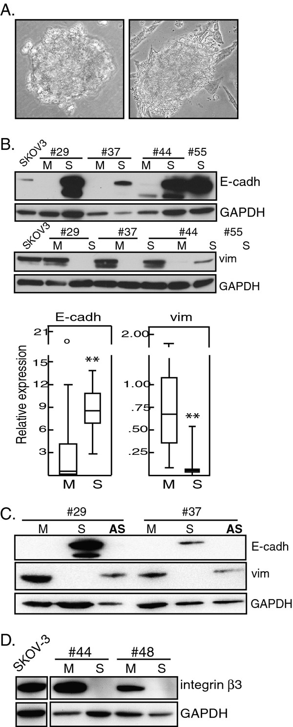
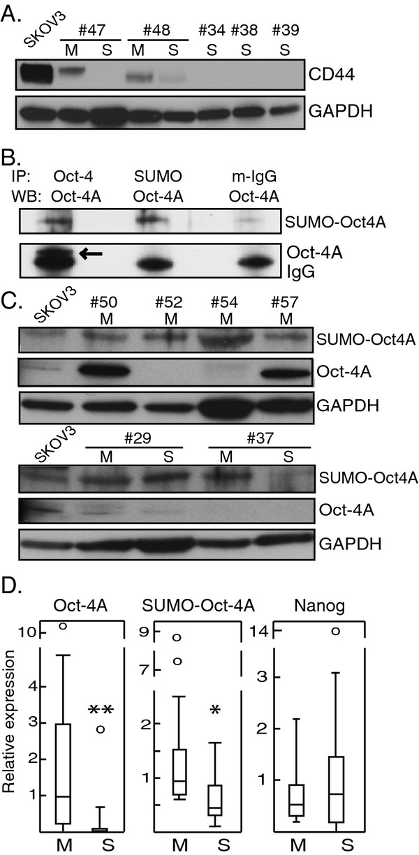
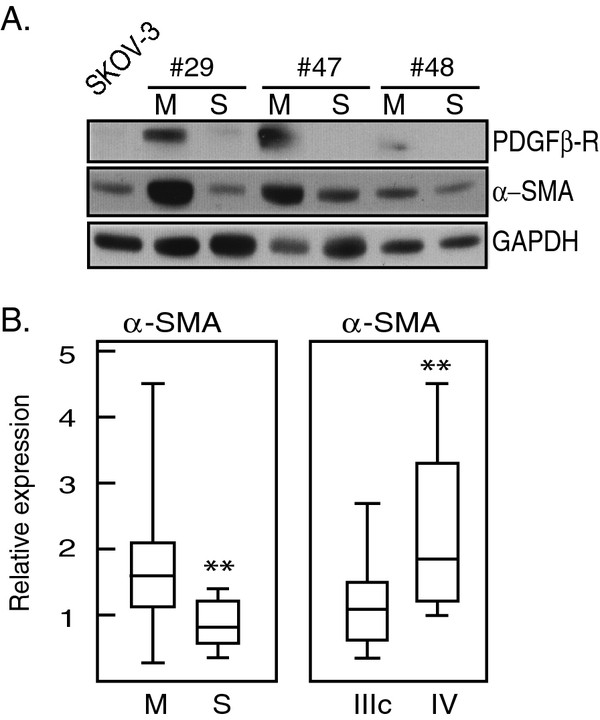
Cells freshly isolated from malignant ascites were separated into sphere samples (S-type samples, n=9) and monolayer-forming single-cell suspensions (M-type, n=18). Using western blot, these were then compared for expression of protein markers of EMT, TIC, and of cancer-associated fibroblasts (CAFs).

RESULTS

S-type cells differed significantly from M-type by expressing high levels of E-cadherin and no or little vimentin, integrin-β3 or stem cell transcription factor Oct-4A. By contrast, M-type samples were enriched for CD44, Oct-4A and for CAF markers. Independently of M- and S-type, there was a strong correlation between TIC markers Nanog and EpCAM. The CAF marker α-SMA correlated with clinical stage IV. This is the first report on CAF markers in malignant ascites and on SUMOylation of Oct-4A in ovarian cancer.

CONCLUSIONS

In addition to demonstrating potentially high levels of TICs in ascites, the results suggest that the S-type population is the less tumorigenic one. Nanog(high)/EpCAM(high) samples represent a TIC subset which may be either M- or S-type, and which is separate from the CD44(high)/Oct-4A(high) subset observed only in M-type samples. This demonstrates a heterogeneity in TIC populations in vivo which has practical implications for TIC isolation based on cell sorting. The biological heterogeneity will need to be addressed in future therapeutical strategies.

摘要

背景

在卵巢癌中,大量的腹腔内播散是由于腹腔内脱落的肿瘤细胞所致。肿瘤起始细胞(TIC 或癌症干细胞)和表现出上皮间质转化(EMT)的细胞尤其与此相关。有时会观察到自发形成的球形细胞聚集,但尽管与体外 TIC 形成的细胞相似,但它们的意义尚不清楚。

方法

从恶性腹腔积液中分离的新鲜细胞被分离成球体样本(S 型样本,n=9)和单层形成的单细胞悬液(M 型,n=18)。然后通过 Western blot 比较这些样本中 EMT、TIC 和癌症相关成纤维细胞(CAF)的蛋白标志物表达。

结果

S 型细胞与 M 型细胞显著不同,表现为高水平的 E-钙黏蛋白,而无或很少表达波形蛋白、整合素-β3 或转录因子 Oct-4A。相比之下,M 型样本富含 CD44、Oct-4A 和 CAF 标志物。与 M-和 S-型无关,TIC 标志物 Nanog 和 EpCAM 之间存在强烈相关性。CAF 标志物α-SMA 与临床分期 IV 相关。这是首次报道恶性腹腔积液中的 CAF 标志物和卵巢癌中 Oct-4A 的 SUMO 化。

结论

除了证明腹水 TIC 水平可能较高外,结果表明 S 型细胞群体的致瘤性较低。Nanog(高)/EpCAM(高)样本代表了一个 TIC 亚群,它可能是 M 型或 S 型,与仅在 M 型样本中观察到的 CD44(高)/Oct-4A(高)亚群不同。这证明了体内 TIC 群体的异质性,这对基于细胞分选的 TIC 分离具有实际意义。在未来的治疗策略中,需要解决这种生物学异质性。

https://cdn.ncbi.nlm.nih.gov/pmc/blobs/fda6/3517779/4b11a7d2ad82/1471-2407-12-359-3.jpg
https://cdn.ncbi.nlm.nih.gov/pmc/blobs/fda6/3517779/5155e7805400/1471-2407-12-359-1.jpg
https://cdn.ncbi.nlm.nih.gov/pmc/blobs/fda6/3517779/2d31986d5443/1471-2407-12-359-2.jpg
https://cdn.ncbi.nlm.nih.gov/pmc/blobs/fda6/3517779/4b11a7d2ad82/1471-2407-12-359-3.jpg
https://cdn.ncbi.nlm.nih.gov/pmc/blobs/fda6/3517779/5155e7805400/1471-2407-12-359-1.jpg
https://cdn.ncbi.nlm.nih.gov/pmc/blobs/fda6/3517779/2d31986d5443/1471-2407-12-359-2.jpg
https://cdn.ncbi.nlm.nih.gov/pmc/blobs/fda6/3517779/4b11a7d2ad82/1471-2407-12-359-3.jpg

相似文献

1
Protein markers of cancer-associated fibroblasts and tumor-initiating cells reveal subpopulations in freshly isolated ovarian cancer ascites.癌症相关成纤维细胞和肿瘤起始细胞的蛋白标志物揭示了新鲜分离的卵巢癌腹水中的亚群。
BMC Cancer. 2012 Aug 18;12:359. doi: 10.1186/1471-2407-12-359.
2
Oct-4 and Nanog promote the epithelial-mesenchymal transition of breast cancer stem cells and are associated with poor prognosis in breast cancer patients.八聚体转录因子4(Oct-4)和Nanog促进乳腺癌干细胞的上皮-间质转化,并与乳腺癌患者的不良预后相关。
Oncotarget. 2014 Nov 15;5(21):10803-15. doi: 10.18632/oncotarget.2506.
3
Different Effects of BORIS/CTCFL on Stemness Gene Expression, Sphere Formation and Cell Survival in Epithelial Cancer Stem Cells.BORIS/CTCFL对上皮癌干细胞干性基因表达、成球能力及细胞存活的不同影响
PLoS One. 2015 Jul 17;10(7):e0132977. doi: 10.1371/journal.pone.0132977. eCollection 2015.
4
Lgr5+CD44+EpCAM+ Strictly Defines Cancer Stem Cells in Human Colorectal Cancer.Lgr5+CD44+EpCAM+严格定义了人类结直肠癌中的癌症干细胞。
Cell Physiol Biochem. 2018;46(2):860-872. doi: 10.1159/000488743. Epub 2018 Mar 29.
5
Isolation and characterization of tumor cells from the ascites of ovarian cancer patients: molecular phenotype of chemoresistant ovarian tumors.从卵巢癌患者腹水中分离和鉴定肿瘤细胞:耐药性卵巢肿瘤的分子表型。
PLoS One. 2012;7(10):e46858. doi: 10.1371/journal.pone.0046858. Epub 2012 Oct 8.
6
Loss of Krüppel-like factor 9 facilitates stemness in ovarian cancer ascites-derived multicellular spheroids via Notch1/slug signaling.Krüppel 样因子 9 的缺失通过 Notch1/slug 信号通路促进卵巢癌腹水来源的多细胞球体的干性。
Cancer Sci. 2021 Oct;112(10):4220-4233. doi: 10.1111/cas.15100. Epub 2021 Aug 20.
7
Mullerian inhibiting substance preferentially inhibits stem/progenitors in human ovarian cancer cell lines compared with chemotherapeutics.Mullerian 抑制物质与人卵巢癌细胞系中的化疗药物相比,优先抑制干细胞/祖细胞。
Proc Natl Acad Sci U S A. 2010 Nov 2;107(44):18874-9. doi: 10.1073/pnas.1012667107. Epub 2010 Oct 15.
8
Microparticles from ovarian carcinomas are shed into ascites and promote cell migration.卵巢癌细胞释放的微颗粒进入腹水并促进细胞迁移。
Int J Gynecol Cancer. 2012 May;22(4):546-52. doi: 10.1097/IGC.0b013e318241d9b9.
9
EMT and acquisition of stem cell-like properties are involved in spontaneous formation of tumorigenic hybrids between lung cancer and bone marrow-derived mesenchymal stem cells.上皮-间质转化(EMT)以及干细胞样特性的获得参与了肺癌与骨髓间充质干细胞之间致瘤性杂交瘤的自发形成。
PLoS One. 2014 Feb 6;9(2):e87893. doi: 10.1371/journal.pone.0087893. eCollection 2014.
10
Notch-1 induces epithelial-mesenchymal transition consistent with cancer stem cell phenotype in pancreatic cancer cells.Notch-1 诱导胰腺癌细胞发生上皮间质转化,符合癌症干细胞表型。
Cancer Lett. 2011 Aug 1;307(1):26-36. doi: 10.1016/j.canlet.2011.03.012. Epub 2011 Apr 3.

引用本文的文献

1
What is the Reason That the Pharmacological Future of Chemotherapeutics in the Treatment of Lung Cancer Could Be Most Closely Related to Nanostructures? Platinum Drugs in Therapy of Non-Small and Small Cell Lung Cancer and Their Unexpected, Possible Interactions. The Review.为什么化疗药物在肺癌治疗中的药理前景可能与纳米结构最密切相关?铂类药物在非小细胞肺癌和小细胞肺癌治疗中的作用及其意想不到的可能相互作用。综述。
Int J Nanomedicine. 2024 Sep 14;19:9503-9547. doi: 10.2147/IJN.S469217. eCollection 2024.
2
Metastasis-associated fibroblasts in peritoneal surface malignancies.腹膜表面恶性肿瘤中的转移相关成纤维细胞。
Br J Cancer. 2024 Aug;131(3):407-419. doi: 10.1038/s41416-024-02717-4. Epub 2024 May 23.
3

本文引用的文献

1
Differentiation of breast cancer stem cells by knockdown of CD44: promising differentiation therapy.敲低 CD44 对乳腺癌干细胞的分化作用:有前景的分化治疗。
J Transl Med. 2011 Dec 7;9:209. doi: 10.1186/1479-5876-9-209.
2
Fibroblasts in omentum activated by tumor cells promote ovarian cancer growth, adhesion and invasiveness.肿瘤细胞激活的大网膜成纤维细胞促进卵巢癌细胞生长、黏附和侵袭。
Carcinogenesis. 2012 Jan;33(1):20-9. doi: 10.1093/carcin/bgr230. Epub 2011 Oct 21.
3
Lessons from common markers of tumor-initiating cells in solid cancers.
Deciphering resistance mechanisms and novel strategies to overcome drug resistance in ovarian cancer: a comprehensive review.
解析卵巢癌耐药机制及克服耐药策略的研究进展:综述
Oncol Res. 2024 Apr 23;32(5):831-847. doi: 10.32604/or.2024.031006. eCollection 2024.
4
Cancer-associated fibroblasts in gynecological malignancies: are they really allies of the enemy?妇科恶性肿瘤中的癌症相关成纤维细胞:它们真的是敌人的盟友吗?
Front Oncol. 2023 Apr 24;13:1106757. doi: 10.3389/fonc.2023.1106757. eCollection 2023.
5
Integrated single-cell and bulk RNA sequencing analyses reveal a prognostic signature of cancer-associated fibroblasts in head and neck squamous cell carcinoma.整合单细胞和批量RNA测序分析揭示头颈部鳞状细胞癌中癌症相关成纤维细胞的预后特征。
Front Genet. 2022 Dec 8;13:1028469. doi: 10.3389/fgene.2022.1028469. eCollection 2022.
6
Spheroid Formation and Peritoneal Metastasis in Ovarian Cancer: The Role of Stromal and Immune Components.卵巢癌球体形成和腹膜转移:基质和免疫成分的作用。
Int J Mol Sci. 2022 Jun 1;23(11):6215. doi: 10.3390/ijms23116215.
7
Matrix stiffening and acquired resistance to chemotherapy: concepts and clinical significance.基质僵硬和获得性化疗耐药:概念和临床意义。
Br J Cancer. 2022 May;126(9):1253-1263. doi: 10.1038/s41416-021-01680-8. Epub 2022 Feb 5.
8
Cancer-associated fibroblast-derived CCL5 contributes to cisplatin resistance in A549 NSCLC cells partially through upregulation of lncRNA HOTAIR expression.癌症相关成纤维细胞衍生的CCL5部分通过上调lncRNA HOTAIR的表达促进A549非小细胞肺癌细胞对顺铂的耐药性。
Oncol Lett. 2021 Oct;22(4):696. doi: 10.3892/ol.2021.12957. Epub 2021 Aug 2.
9
Loss of Krüppel-like factor 9 facilitates stemness in ovarian cancer ascites-derived multicellular spheroids via Notch1/slug signaling.Krüppel 样因子 9 的缺失通过 Notch1/slug 信号通路促进卵巢癌腹水来源的多细胞球体的干性。
Cancer Sci. 2021 Oct;112(10):4220-4233. doi: 10.1111/cas.15100. Epub 2021 Aug 20.
10
Fibroblasts in cancer dormancy: foe or friend?癌症休眠中的成纤维细胞:敌还是友?
Cancer Cell Int. 2021 Mar 26;21(1):184. doi: 10.1186/s12935-021-01883-2.
实体瘤中肿瘤起始细胞的常见标志物带来的启示。
Cell Mol Life Sci. 2011 Dec;68(24):4009-22. doi: 10.1007/s00018-011-0772-9. Epub 2011 Jul 24.
4
Integrin regulation of beta-catenin signaling in ovarian carcinoma.整合素在卵巢癌中对β-连环蛋白信号的调节。
J Biol Chem. 2011 Jul 1;286(26):23467-75. doi: 10.1074/jbc.M110.199539. Epub 2011 Apr 25.
5
Expression of aldehyde dehydrogenase and CD133 defines ovarian cancer stem cells.醛脱氢酶和 CD133 的表达定义了卵巢癌干细胞。
Int J Cancer. 2012 Jan 1;130(1):29-39. doi: 10.1002/ijc.25967. Epub 2011 Apr 8.
6
Inconsistent immunohistochemical expression patterns of four different CD133 antibody clones in glioblastoma.胶质母细胞瘤中四种不同 CD133 抗体克隆的免疫组化表达模式不一致。
J Histochem Cytochem. 2011 Apr;59(4):391-407. doi: 10.1369/0022155411400867.
7
Ovarian cancer-associated fibroblasts contribute to epithelial ovarian carcinoma metastasis by promoting angiogenesis, lymphangiogenesis and tumor cell invasion.卵巢癌相关成纤维细胞通过促进血管生成、淋巴管生成和肿瘤细胞侵袭促进上皮性卵巢癌转移。
Cancer Lett. 2011 Apr 1;303(1):47-55. doi: 10.1016/j.canlet.2011.01.011.
8
Nanog is highly expressed in ovarian serous cystadenocarcinoma and correlated with clinical stage and pathological grade.Nanog 在卵巢浆液性囊腺癌中高表达,与临床分期和病理分级相关。
Pathobiology. 2010;77(6):283-8. doi: 10.1159/000320866. Epub 2011 Jan 24.
9
Analysis of epithelial and mesenchymal markers in ovarian cancer reveals phenotypic heterogeneity and plasticity.分析卵巢癌中的上皮和间充质标志物揭示了表型异质性和可塑性。
PLoS One. 2011 Jan 14;6(1):e16186. doi: 10.1371/journal.pone.0016186.
10
Expression of octamer-4 in serous and mucinous ovarian carcinoma.Octamer-4 在浆液性和黏液性卵巢癌中的表达。
J Clin Pathol. 2010 Oct;63(10):879-83. doi: 10.1136/jcp.2009.073593.