Mavromatis Kreton, Sutcliffe Diane J, Joseph Giji, Alexander R Wayne, Waller Edmund K, Quyyumi Arshed A, Taylor W Robert
Department of Medicine, Emory University, Atlanta, GA, USA.
J Biomol Screen. 2012 Oct;17(9):1128-35. doi: 10.1177/1087057112457043. Epub 2012 Aug 17.
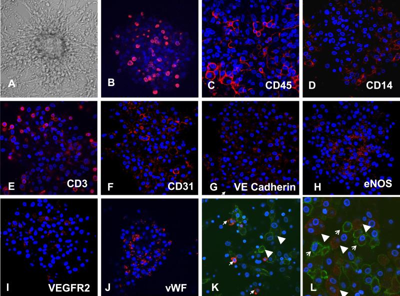
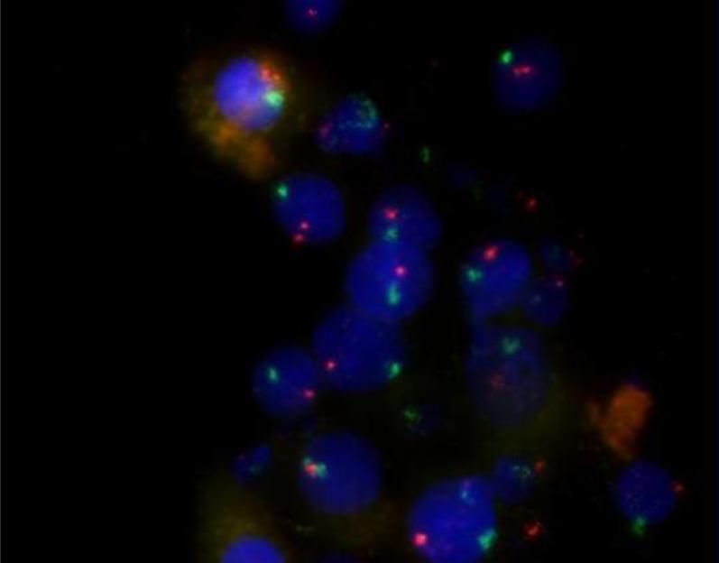
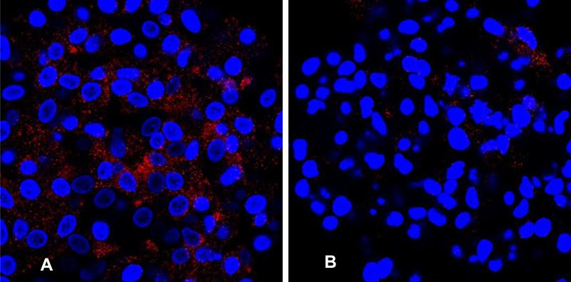
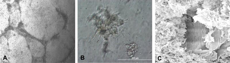
Although multiple culture assays have been designed to identify endothelial progenitor cells (EPCs), the phenotype of cells grown in culture often remains undefined. We sought to define and characterize the proangiogenic cell population within human peripheral blood mononuclear cells. Mononuclear cells were isolated from peripheral blood and grown under angiogenic conditions for 7 days. Formed colonies (CFU-As) were identified and analyzed for proliferation, mRNA and surface antigen expression, tube-forming ability, and chromosomal content. Colonies were composed of a heterogeneous group of cells expressing the leukocyte antigens CD45, CD14, and CD3, as well as the endothelial proteins vascular endothelial (VE) cadherin, von Willebrand's factor (vWF), CD31, and endothelial nitric oxide synthase (eNOS). Colony cells expressed increased levels of proangiogenic growth factors, and they formed tubes in Matrigel. In comparison with colonies from the CFU-Hill assay, our assay resulted in a greater number of colonies (19 ± 9 vs. 13 ± 7; p < 0.0001) with a substantial number of cells expressing an endothelial phenotype (20.2% ± 7.4% vs. 2.2% ± 1.2% expressing eNOS, p = 0.0006). Chromosomal analysis indicated the colony cells were bone marrow derived. We, therefore, describe a colony-forming unit assay that measures bone marrow-derived circulating mononuclear cells with the capacity to proliferate and mature into proangiogenic leukocytic and endothelial-like cells. This assay, therefore, reflects circulating, bone marrow-derived proangiogenic activity.
尽管已经设计了多种培养分析方法来鉴定内皮祖细胞(EPC),但培养中生长的细胞表型往往仍不明确。我们试图定义和表征人类外周血单个核细胞中的促血管生成细胞群。从外周血中分离出单个核细胞,并在促血管生成条件下培养7天。鉴定形成的集落(CFU-A),并分析其增殖、mRNA和表面抗原表达、管形成能力及染色体含量。集落由一群异质性细胞组成,这些细胞表达白细胞抗原CD45、CD14和CD3,以及内皮蛋白血管内皮(VE)钙黏蛋白、血管性血友病因子(vWF)、CD31和内皮型一氧化氮合酶(eNOS)。集落细胞表达促血管生成生长因子的水平增加,并且它们在基质胶中形成管。与CFU-Hill分析中的集落相比,我们的分析产生了更多的集落(19±9比13±7;p<0.0001),有大量细胞表达内皮表型(表达eNOS的细胞为20.2%±7.4%比2.2%±1.2%,p = 0.0006)。染色体分析表明集落细胞来源于骨髓。因此,我们描述了一种集落形成单位分析方法,该方法可测量具有增殖并成熟为促血管生成白细胞样和内皮样细胞能力的骨髓来源循环单个核细胞。因此,该分析反映了循环的、骨髓来源的促血管生成活性。