Bone Structural Biology Laboratory, Faculty of Biological Sciences, University of Leeds , Leeds, Yorkshire, UK.
Front Endocrinol (Lausanne). 2012 Aug 9;3:98. doi: 10.3389/fendo.2012.00098. eCollection 2012.
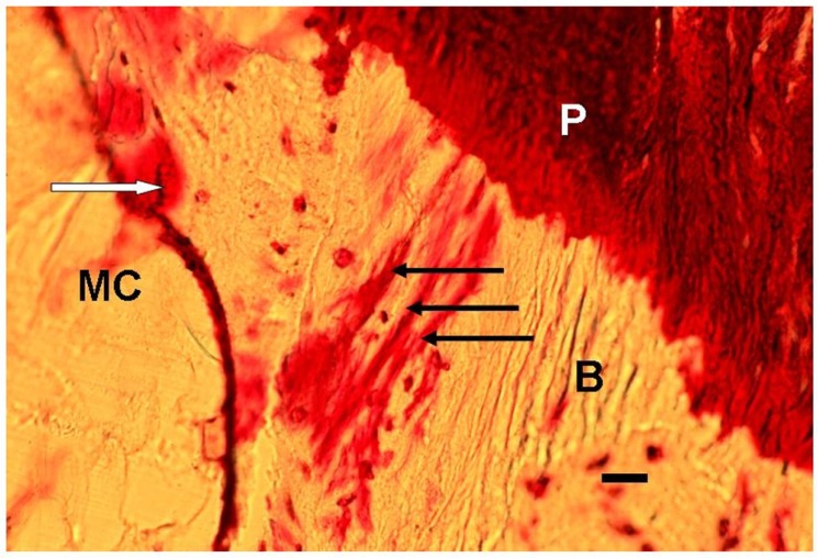
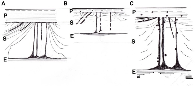
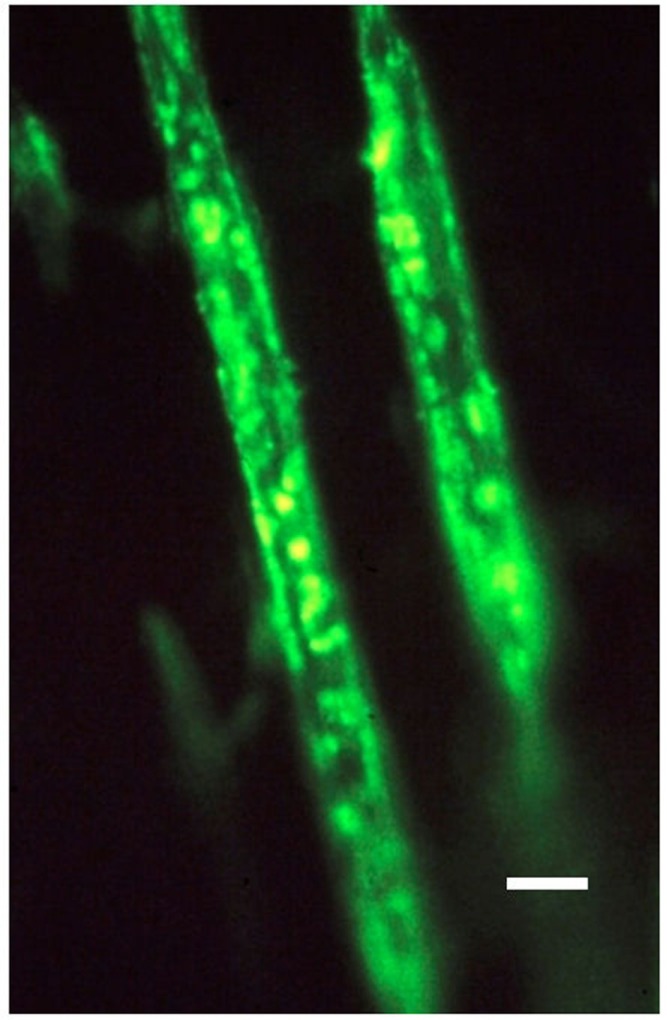
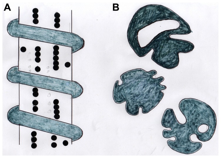
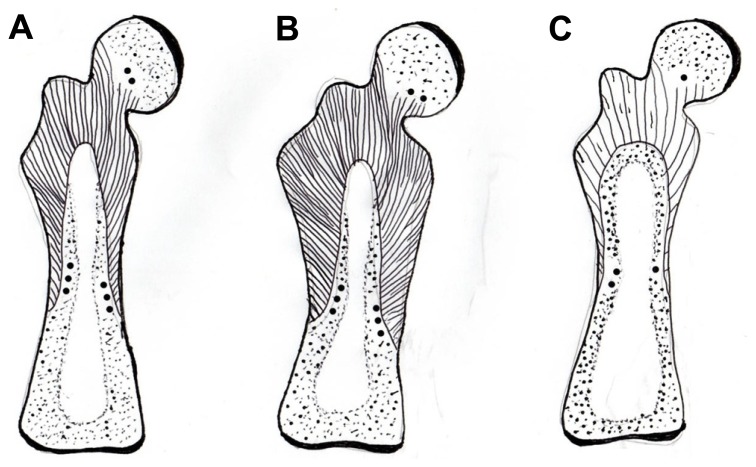
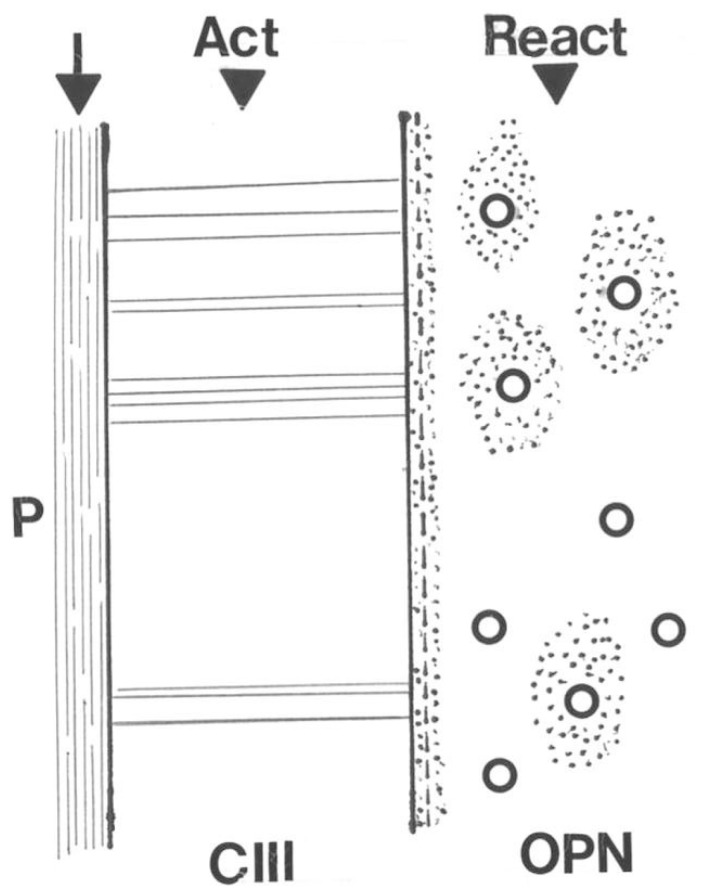
Sharpey's "perforating" fibers (SF) are well known skeletally in tooth anchorage. Elsewhere they provide anchorage for the periosteum and are less well documented. Immunohistochemistry has transformed their potential significance by identifying their collagen type III (CIII) content and enabling their mapping in domains as permeating arrays of fibers (5-25 μ thick), protected from osteoclastic resorption by their poor mineralization. As periosteal extensions they are crucial in early skeletal development and central to intramembranous bone healing, providing unique microanatomical avenues for musculoskeletal exchange, their composition (e.g., collagen type VI, elastin, tenascin) combined with a multiaxial pattern of insertion suggesting a role more complex than attachment alone would justify. A proportion permeate the cortex to the endosteum (and beyond), fusing into a CIII-rich osteoid layer (<2 μ thick) encompassing all resting surfaces, and with which they apparently integrate into a PERIOSTEAL-SHARPEY FIBER-ENDOSTEUM (PSE) structural continuum. This intraosseous system behaves in favor of bone loss or gain depending upon extraneous stimuli (i.e., like Frost's hypothetical "mechanostat"). Thus, the birefringent fibers are sensitive to humoral factors (e.g., estrogen causes retraction, rat femur model), physical activity (e.g., running causes expansion, rat model), aging (e.g., causes fragmentation, pig mandible model), and pathology (e.g., atrophied in osteoporosis, hypertrophied in osteoarthritis, human proximal femur), and with encroaching mineral particles hardening the usually soft parts. In this way the unobtrusive periosteal SF network may regulate bone status, perhaps even contributing to predictable "hotspots" of trabecular disconnection, particularly at sites of tension prone to fatigue, and with the network deteriorating significantly before bone matrix loss.
Sharpey 的“穿孔”纤维(SF)在牙齿锚固中在骨骼中广为人知。在其他地方,它们为骨膜提供锚固,并且记录较少。免疫组织化学通过鉴定其 III 型胶原(CIII)含量并能够在穿透纤维阵列(5-25μm 厚)的域中对其进行映射,从而改变了其潜在意义,这些纤维通过其较差的矿化而免受破骨细胞吸收的保护。作为骨膜延伸,它们在早期骨骼发育中至关重要,并且是膜内骨愈合的核心,为肌肉骨骼交换提供了独特的微观解剖途径,其组成(例如,胶原 VI 型,弹性蛋白,腱蛋白)结合多轴插入模式表明其作用比单独附着更复杂。一部分渗透到皮质到骨内膜(甚至更远),融合到富含 CIII 的类骨质层(<2μm 厚),包含所有静止表面,并且它们显然与骨内膜 - Sharpey 纤维 - 骨内膜(PSE)结构连续体整合在一起。这个骨内系统根据外来刺激(即类似于 Frost 的假设“机械感受器”)表现为有利于骨质流失或增加。因此,双折射纤维对体液因子敏感(例如,雌激素导致收缩,大鼠股骨模型),身体活动(例如,跑步导致扩张,大鼠模型),衰老(例如,导致碎片化,猪下颌骨模型),和病理学(例如,骨质疏松症中萎缩,骨关节炎中肥大,人近端股骨),并且随着侵入性矿物质颗粒的增加,通常较软的部分变硬。通过这种方式,不起眼的骨膜 SF 网络可以调节骨骼状态,甚至可能有助于预测小梁分离的“热点”,尤其是在容易疲劳的张力部位,并且在骨基质丢失之前,网络会明显恶化。