Bellvitge Biomedical Research Institute, IDIBELL, Barcelona, Spain.
PLoS One. 2012;7(8):e42994. doi: 10.1371/journal.pone.0042994. Epub 2012 Aug 13.
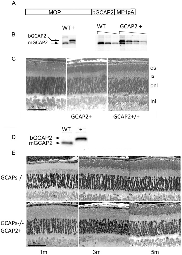
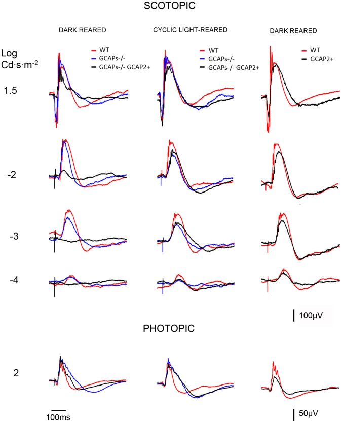
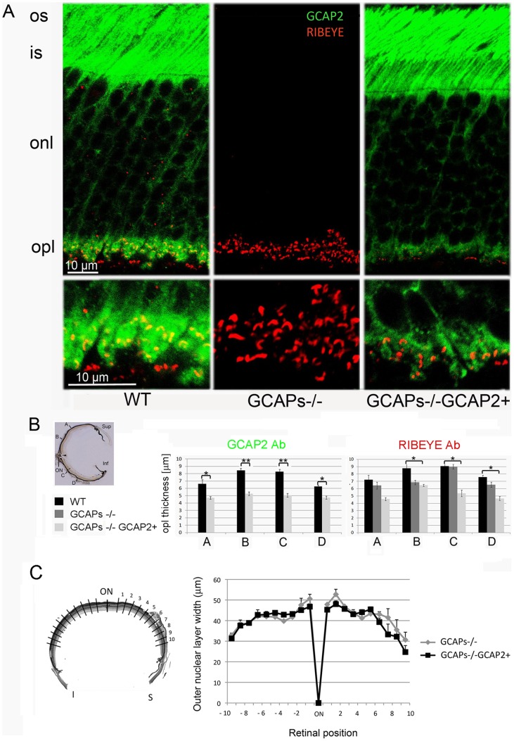
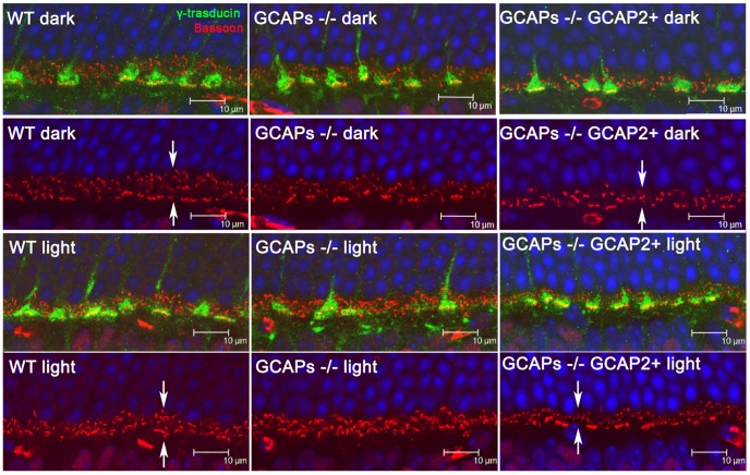
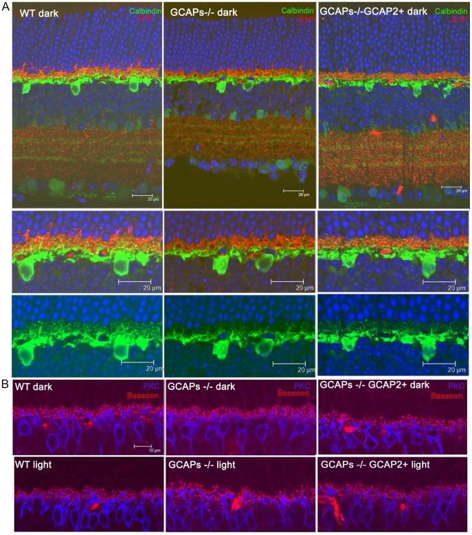
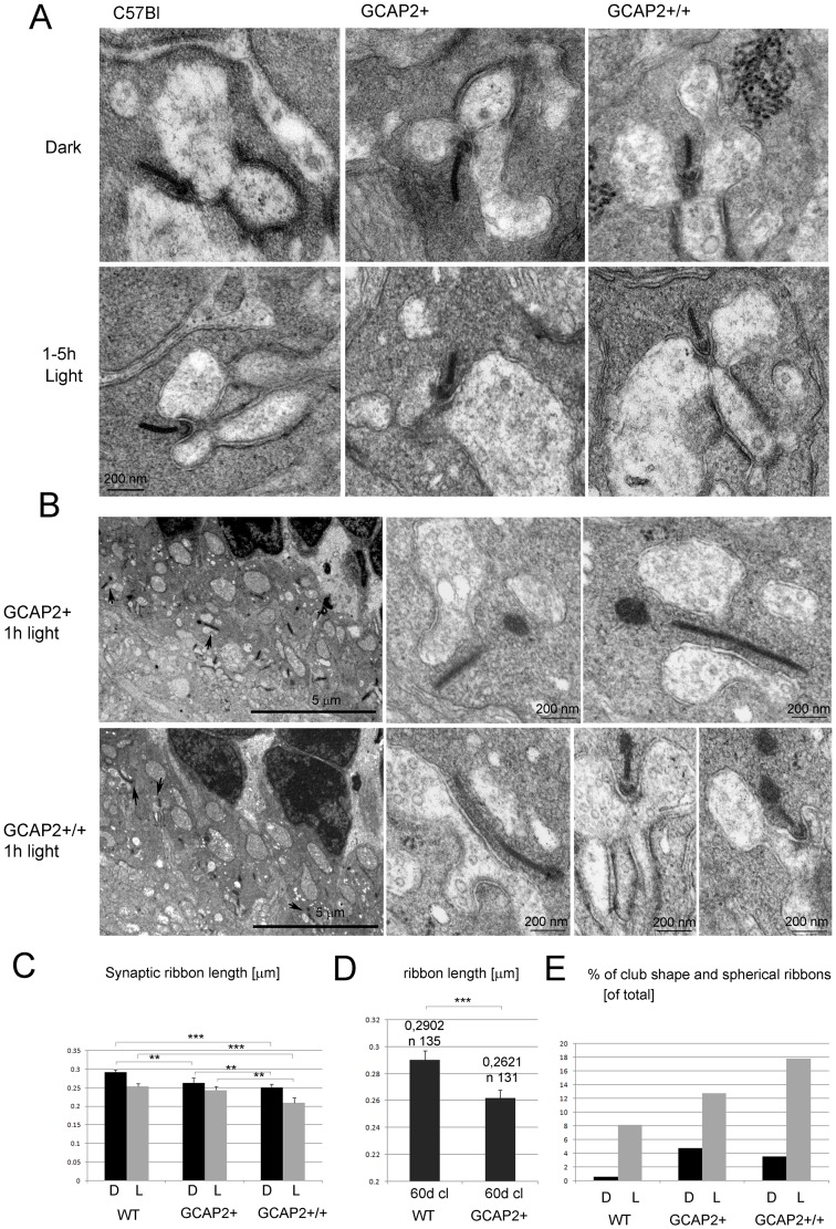
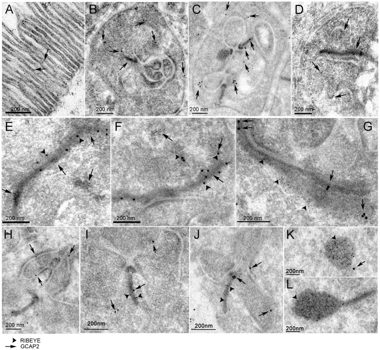
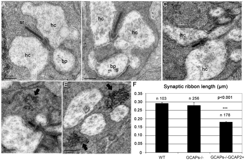
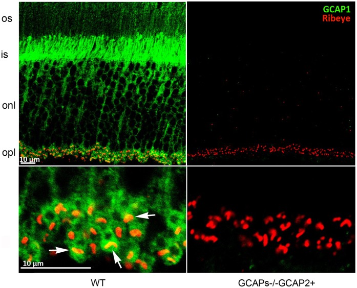
Guanylate cyclase activating proteins are EF-hand containing proteins that confer calcium sensitivity to retinal guanylate cyclase at the outer segment discs of photoreceptor cells. By making the rate of cGMP synthesis dependent on the free intracellular calcium levels set by illumination, GCAPs play a fundamental role in the recovery of the light response and light adaptation. The main isoforms GCAP1 and GCAP2 also localize to the synaptic terminal, where their function is not known. Based on the reported interaction of GCAP2 with Ribeye, the major component of synaptic ribbons, it was proposed that GCAP2 could mediate the synaptic ribbon dynamic changes that happen in response to light. We here present a thorough ultrastructural analysis of rod synaptic terminals in loss-of-function (GCAP1/GCAP2 double knockout) and gain-of-function (transgenic overexpression) mouse models of GCAP2. Rod synaptic ribbons in GCAPs-/- mice did not differ from wildtype ribbons when mice were raised in constant darkness, indicating that GCAPs are not required for ribbon early assembly or maturation. Transgenic overexpression of GCAP2 in rods led to a shortening of synaptic ribbons, and to a higher than normal percentage of club-shaped and spherical ribbon morphologies. Restoration of GCAP2 expression in the GCAPs-/- background (GCAP2 expression in the absence of endogenous GCAP1) had the striking result of shortening ribbon length to a much higher degree than overexpression of GCAP2 in the wildtype background, as well as reducing the thickness of the outer plexiform layer without affecting the number of rod photoreceptor cells. These results indicate that preservation of the GCAP1 to GCAP2 relative levels is relevant for maintaining the integrity of the synaptic terminal. Our demonstration of GCAP2 immunolocalization at synaptic ribbons at the ultrastructural level would support a role of GCAPs at mediating the effect of light on morphological remodeling changes of synaptic ribbons.
鸟苷酸环化酶激活蛋白是 EF 手型结构域蛋白,可在外节盘状结构赋予光感受器细胞中的视网膜鸟苷酸环化酶对钙离子的敏感性。GCAPs 通过使 cGMP 合成的速率依赖于光照设定的细胞内游离钙离子水平,从而在光反应和光适应的恢复中发挥基本作用。主要同工型 GCAP1 和 GCAP2 也定位于突触末端,但它们的功能尚不清楚。基于已报道的 GCAP2 与突触小带主要成分 Ribeye 的相互作用,提出 GCAP2 可能介导光响应时发生的突触小带动态变化。我们在此报告了 GCAP2 功能丧失(GCAP1/GCAP2 双敲除)和功能获得(转基因过表达)小鼠模型中的杆状突触末端的详细超微结构分析。当小鼠在持续黑暗中生长时,GCAPs-/- 小鼠中的杆状突触小带与野生型小带没有差异,这表明 GCAPs 对于小带的早期组装或成熟不是必需的。GCAP2 在杆状细胞中的转基因过表达导致突触小带缩短,并导致高于正常比例的棒状和球形小带形态。在 GCAPs-/- 背景中恢复 GCAP2 的表达(在缺乏内源性 GCAP1 的情况下表达 GCAP2)产生了惊人的结果,即与在野生型背景中过表达 GCAP2 相比,小带长度缩短的程度更高,并且外丛状层的厚度减小而不影响杆状光感受器细胞的数量。这些结果表明,保持 GCAP1 与 GCAP2 的相对水平对于维持突触末端的完整性是相关的。我们在超微结构水平上证明了 GCAP2 在突触小带上的免疫定位,这支持了 GCAPs 在介导光对突触小带形态重塑变化的影响中的作用。