Department of Neurosciences, Biomedicine and Movement Sciences, Section of Biological Chemistry, University of Verona, 37129 Verona, Italy.
MAGI'S Lab S.R.L., 38068 Rovereto, Italy.
Int J Mol Sci. 2021 Apr 14;22(8):4030. doi: 10.3390/ijms22084030.
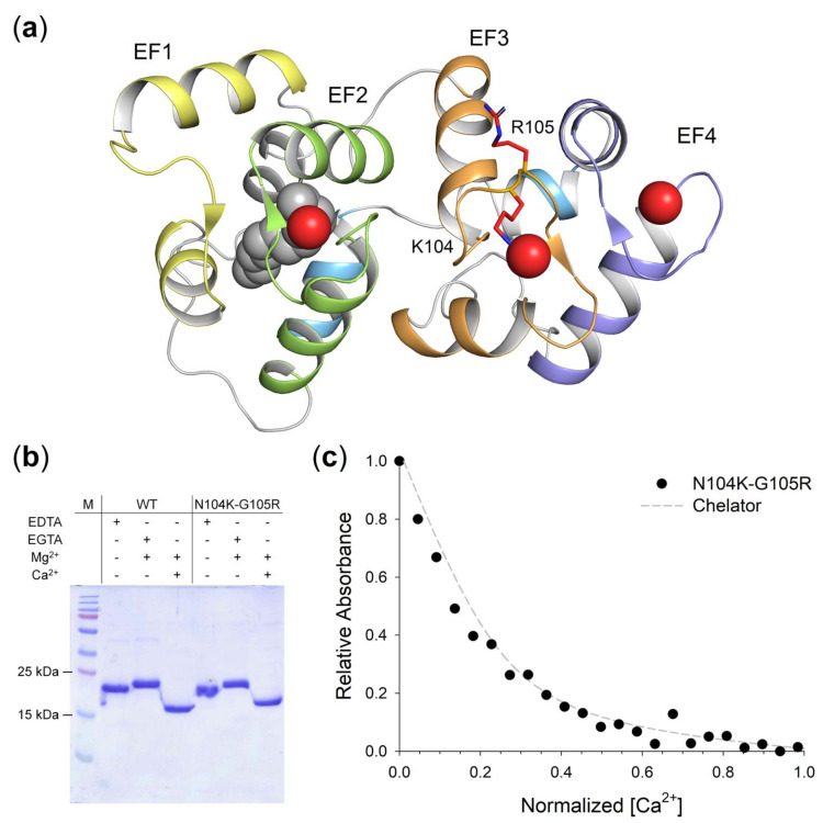
Guanylate cyclase-activating protein 1 (GCAP1) is involved in the shutdown of the phototransduction cascade by regulating the enzymatic activity of retinal guanylate cyclase via a Ca/cGMP negative feedback. While the phototransduction-associated role of GCAP1 in the photoreceptor outer segment is widely established, its implication in synaptic transmission to downstream neurons remains to be clarified. Here, we present clinical and biochemical data on a novel isolate GCAP1 variant leading to a double amino acid substitution (p.N104K and p.G105R) and associated with cone dystrophy (COD) with an unusual phenotype. Severe alterations of the electroretinogram were observed under both scotopic and photopic conditions, with a negative pattern and abnormally attenuated b-wave component. The biochemical and biophysical analysis of the heterologously expressed N104K-G105R variant corroborated by molecular dynamics simulations highlighted a severely compromised Ca-sensitivity, accompanied by minor structural and stability alterations. Such differences reflected on the dysregulation of both guanylate cyclase isoforms (RetGC1 and RetGC2), resulting in the constitutive activation of both enzymes at physiological levels of Ca. As observed with other GCAP1-associated COD, perturbation of the homeostasis of Ca and cGMP may lead to the toxic accumulation of second messengers, ultimately triggering cell death. However, the abnormal electroretinogram recorded in this patient also suggested that the dysregulation of the GCAP1-cyclase complex further propagates to the synaptic terminal, thereby altering the ON-pathway related to the b-wave generation. In conclusion, the pathological phenotype may rise from a combination of second messengers' accumulation and dysfunctional synaptic communication with bipolar cells, whose molecular mechanisms remain to be clarified.
鸟苷酸环化酶激活蛋白 1(GCAP1)通过 Ca/cGMP 负反馈调节视网膜鸟苷酸环化酶的酶活性,参与光转导级联的关闭。虽然 GCAP1 在光感受器外段中的光转导相关作用已得到广泛证实,但它在向下游神经元的突触传递中的作用仍有待阐明。在这里,我们提供了一个新的 GCAP1 变体的临床和生化数据,该变体导致双氨基酸取代(p.N104K 和 p.G105R),并与 Cone 变性(COD)相关,具有不寻常的表型。在暗适应和明适应条件下,都观察到视网膜电图的严重改变,具有负模式和异常衰减的 b 波成分。通过分子动力学模拟对异源表达的 N104K-G105R 变体进行的生化和生物物理分析证实了 Ca 敏感性严重受损,同时伴有微小的结构和稳定性改变。这些差异反映在两种鸟苷酸环化酶同工型(RetGC1 和 RetGC2)的调节失调上,导致在生理 Ca 水平下两种酶的组成型激活。与其他 GCAP1 相关的 COD 一样,Ca 和 cGMP 的平衡失调可能导致第二信使的毒性积累,最终触发细胞死亡。然而,在该患者中记录的异常视网膜电图也表明,GCAP1-环化酶复合物的失调进一步传播到突触末端,从而改变与 b 波产生相关的 ON 通路。总之,病理表型可能源于第二信使的积累和与双极细胞的功能失调的突触通讯的组合,其分子机制仍有待阐明。