Department of Radiology and Radiological Science, Medical University of South Carolina, Charleston, SC 29425-3230, USA.
AJR Am J Roentgenol. 2012 Sep;199(3):W392-401. doi: 10.2214/AJR.11.7255.
The purpose of this study was to assess the sensitivities and false-detection rates of two computer-aided detection (CADe) systems when applied to digital or film-screen mammograms in detecting the known breast cancer cases from the Digital Mammographic Imaging Screening Trial (DMIST) breast cancer screening population.
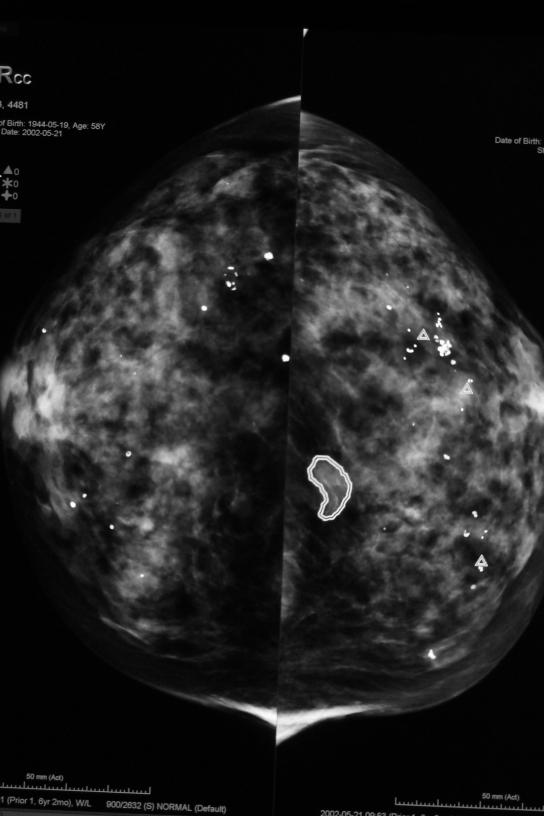
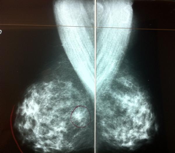
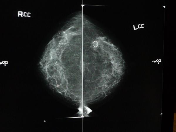
Available film-screen and digital mammograms of 161 breast cancer cases from DMIST were analyzed by two CADe systems, iCAD Second-Look and R2 ImageChecker. Three experienced breast-imaging radiologists reviewed the CADe marks generated for each available cancer case, recording the number and locations of CADe marks and whether each CADe mark location corresponded with the known location of the cancer.
For the 161 cancer cases included in this study, the sensitivities of the DMIST reader without CAD were 0.43 (69/161, 95% CI 0.35-0.51) for digital and 0.41 (66/161, 0.33-0.49) for film-screen mammography. The sensitivities of iCAD were 0.74 (119/161, 0.66-0.81) for digital and 0.69 (111/161, 0.61-0.76) for film-screen mammography, both significantly higher than the DMIST study sensitivities (p < 0.0001 for both). The average number of false CADe marks per case of iCAD was 2.57 (SD, 1.92) for digital and 3.06(1.72) for film-screen mammography. The sensitivity of R2 was 0.74 (119/161, 0.66-0.81) for digital, and 0.60 (97/161, 0.52-0.68) for film-screen mammography, both significantly higher than the DMIST study sensitivities (p < 0.0001 for both). The average number of false CADe marks per case of R2 was 2.07 (1.57) for digital and 1.52 (1.45) for film-screen mammography.
Our results suggest the use of CADe in interpretation of digital and film-screen mammograms could lead to improvements in cancer detection.
本研究旨在评估两种计算机辅助检测(CADe)系统在检测 Digital Mammographic Imaging Screening Trial(DMIST)乳腺癌筛查人群中已知乳腺癌病例时,应用于数字或胶片乳腺 X 线摄影的敏感性和假阳性检出率。
对 DMIST 中 161 例乳腺癌病例的现有胶片和数字乳腺 X 线摄影进行分析,使用 iCAD Second-Look 和 R2 ImageChecker 两种 CADe 系统。三位经验丰富的乳腺影像学放射科医生对每个可用癌症病例生成的 CADe 标记进行了回顾,记录 CADe 标记的数量和位置,以及每个 CADe 标记位置是否与癌症的已知位置相对应。
在本研究中纳入的 161 例癌症病例中,DMIST 无 CAD 读者的敏感性为数字乳腺 X 线摄影 0.43(69/161,95%CI 0.35-0.51),胶片乳腺 X 线摄影 0.41(66/161,0.33-0.49)。iCAD 的敏感性为数字乳腺 X 线摄影 0.74(119/161,0.66-0.81),胶片乳腺 X 线摄影 0.69(111/161,0.61-0.76),均显著高于 DMIST 研究的敏感性(均 p < 0.0001)。iCAD 每个病例的平均假 CADe 标记数为数字乳腺 X 线摄影 2.57(SD,1.92),胶片乳腺 X 线摄影 3.06(1.72)。R2 的敏感性为数字乳腺 X 线摄影 0.74(119/161,0.66-0.81),胶片乳腺 X 线摄影 0.60(97/161,0.52-0.68),均显著高于 DMIST 研究的敏感性(均 p < 0.0001)。R2 每个病例的平均假 CADe 标记数为数字乳腺 X 线摄影 2.07(1.57),胶片乳腺 X 线摄影 1.52(1.45)。
我们的结果表明,在数字和胶片乳腺 X 线摄影的解读中使用 CADe 可能会提高癌症检测的敏感性。