• 文献检索
  • 文档翻译
  • 深度研究
  • 学术资讯
  • Suppr Zotero 插件Zotero 插件
  • 邀请有礼
  • 套餐&价格
  • 历史记录
应用&插件
Suppr Zotero 插件Zotero 插件浏览器插件Mac 客户端Windows 客户端微信小程序
定价
高级版会员购买积分包购买API积分包
服务
文献检索文档翻译深度研究API 文档MCP 服务
关于我们
关于 Suppr公司介绍联系我们用户协议隐私条款
关注我们

Suppr 超能文献

核心技术专利:CN118964589B侵权必究
粤ICP备2023148730 号-1Suppr @ 2026

文献检索

告别复杂PubMed语法,用中文像聊天一样搜索,搜遍4000万医学文献。AI智能推荐,让科研检索更轻松。

立即免费搜索

文件翻译

保留排版,准确专业,支持PDF/Word/PPT等文件格式,支持 12+语言互译。

免费翻译文档

深度研究

AI帮你快速写综述,25分钟生成高质量综述,智能提取关键信息,辅助科研写作。

立即免费体验

墨西哥经过最低限度培训的操作人员利用计算机辅助诊断和低成本超声对可触及乳腺肿块进行分类

Palpable Breast Lump Triage by Minimally Trained Operators in Mexico Using Computer-Assisted Diagnosis and Low-Cost Ultrasound.

作者信息

Love Susan M, Berg Wendie A, Podilchuk Christine, López Aldrete Ana Lilia, Gaxiola Mascareño Aarón Patricio, Pathicherikollamparambil Krishnamohan, Sankarasubramanian Ananth, Eshraghi Leah, Mammone Richard

机构信息

Susan M. Love and Leah Eshraghi, Dr Susan Love Research Foundation, Encino, CA; Wendie A. Berg, Magee-Womens Hospital, University of Pittsburgh School of Medicine, Pittsburgh, PA; Christine Podilchuk, Krishnamohan Pathicherikollamparambil, Ananth Sankarasubramanian, and Richard Mammone, AI Strategy, Warren, NJ; Richard Mammone, Rutgers University, New Brunswick, NJ; and Ana Lilia López Aldrete and Aarón Patricio Gaxiola Mascareño, Instituto de Seguridad y Servicios Sociales de los Trabajadores del Estado Hospital Regional Valentin Gomez Farias, Jalisco, Mexico.

出版信息

J Glob Oncol. 2018 Aug;4:1-9. doi: 10.1200/JGO.17.00222.

DOI:10.1200/JGO.17.00222
PMID:30156946
原文链接:https://pmc.ncbi.nlm.nih.gov/articles/PMC6223536/
Abstract

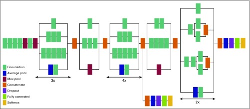
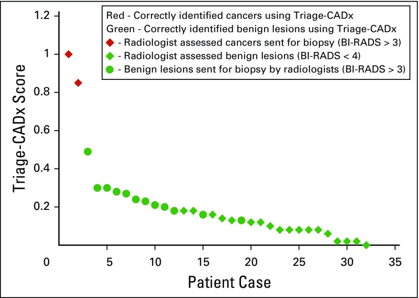
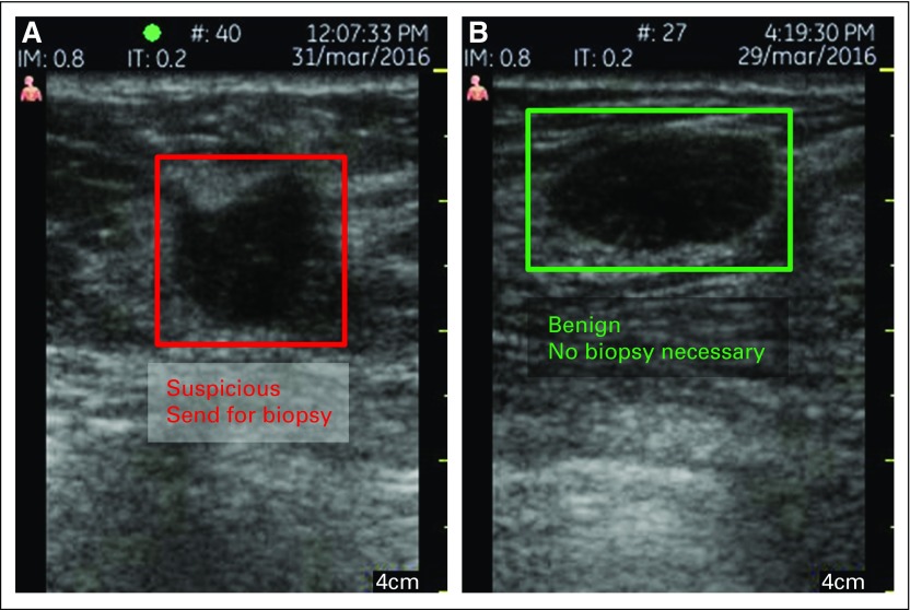
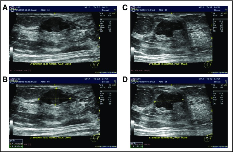
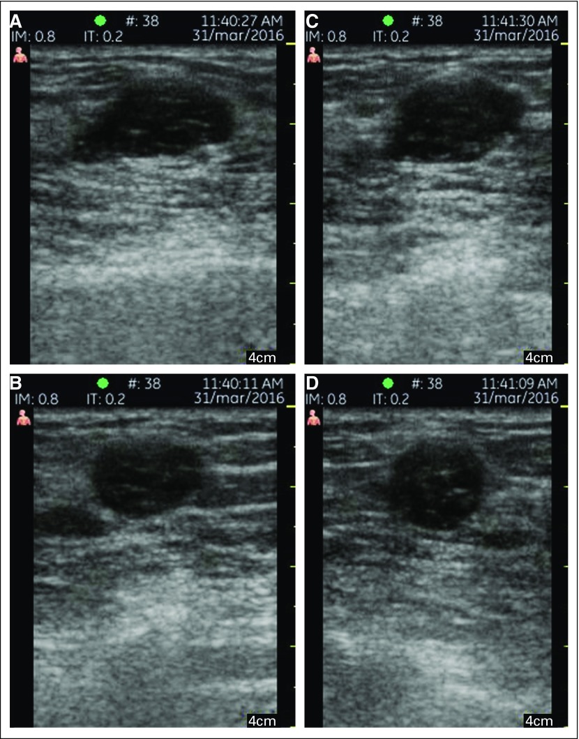
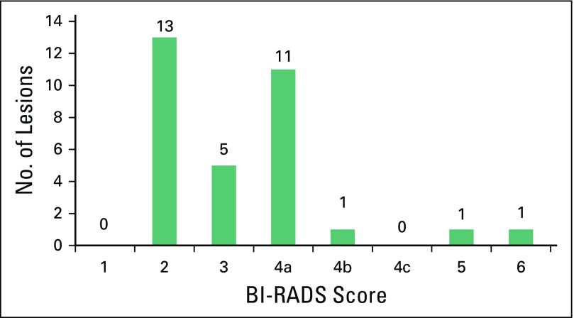
Purpose In low- to middle-income countries (LMICs), most breast cancers present as palpable lumps; however, most palpable lumps are benign. We have developed artificial intelligence-based computer-assisted diagnosis (CADx) for an existing low-cost portable ultrasound system to triage which lumps need further evaluation and which are clearly benign. This pilot study was conducted to demonstrate that this approach can be successfully used by minimally trained health care workers in an LMIC country. Patients and Methods We recruited and trained three nonradiologist health care workers to participate in an institutional review board-approved, Health Insurance Portability and Accountability Act-compliant pilot study in Jalisco, Mexico, to determine whether they could use portable ultrasound (GE Vscan Dual Probe) to acquire images of palpable breast lumps of adequate quality for accurate computer analysis. Images from 32 women with 32 breast masses were then analyzed with a triage-CADx system, generating an output of benign or suspicious (biopsy recommended). Triage-CADx outputs were compared with radiologist readings. Results The nonradiologists were able to acquire adequate images. Triage by the CADx software was as accurate as assessment by specialist radiologists, with two (100%) of two cancers considered suspicious and 30 (100%) of 30 benign lesions classified as benign. Conclusion A portable ultrasound system with CADx software can be successfully used by first-level health care workers to triage palpable breast lumps. These results open up the possibility of implementing practical, cost-effective triage of palpable breast lumps, ensuring that scarce resources can be dedicated to suspicious lesions requiring further workup.

摘要

目的 在低收入和中等收入国家(LMICs),大多数乳腺癌表现为可触及的肿块;然而,大多数可触及的肿块是良性的。我们为现有的低成本便携式超声系统开发了基于人工智能的计算机辅助诊断(CADx),以对哪些肿块需要进一步评估以及哪些明显是良性的进行分类。本试点研究旨在证明这种方法可以被LMIC国家中受过最少培训的医护人员成功使用。

患者与方法 我们招募并培训了三名非放射科医护人员,参与在墨西哥哈利斯科州进行的一项经机构审查委员会批准、符合《健康保险流通与责任法案》的试点研究,以确定他们是否能够使用便携式超声(GE Vscan双探头)获取质量足够的可触及乳腺肿块图像,用于准确的计算机分析。然后使用分类CADx系统分析来自32名患有32个乳腺肿块的女性的图像,生成良性或可疑(建议活检)的输出结果。将分类CADx的输出结果与放射科医生的诊断结果进行比较。

结果 非放射科医生能够获取足够的图像。CADx软件的分类与专科放射科医生的评估一样准确,2个癌症中有2个(100%)被认为可疑,30个良性病变中有30个(100%)被分类为良性。

结论 配备CADx软件的便携式超声系统可以被一级医护人员成功用于对可触及乳腺肿块进行分类。这些结果为实施实用、经济有效的可触及乳腺肿块分类开辟了可能性,确保稀缺资源能够用于需要进一步检查的可疑病变。

https://cdn.ncbi.nlm.nih.gov/pmc/blobs/b829/6223536/de458b3202bd/JGO.17.00222f6.jpg
https://cdn.ncbi.nlm.nih.gov/pmc/blobs/b829/6223536/740deb67e8b4/JGO.17.00222f1.jpg
https://cdn.ncbi.nlm.nih.gov/pmc/blobs/b829/6223536/fb986ebf861e/JGO.17.00222f2.jpg
https://cdn.ncbi.nlm.nih.gov/pmc/blobs/b829/6223536/a7f72d709afe/JGO.17.00222f3.jpg
https://cdn.ncbi.nlm.nih.gov/pmc/blobs/b829/6223536/55eb154f1337/JGO.17.00222f4.jpg
https://cdn.ncbi.nlm.nih.gov/pmc/blobs/b829/6223536/89db9885085e/JGO.17.00222f5.jpg
https://cdn.ncbi.nlm.nih.gov/pmc/blobs/b829/6223536/de458b3202bd/JGO.17.00222f6.jpg

相似文献

1
Palpable Breast Lump Triage by Minimally Trained Operators in Mexico Using Computer-Assisted Diagnosis and Low-Cost Ultrasound.墨西哥经过最低限度培训的操作人员利用计算机辅助诊断和低成本超声对可触及乳腺肿块进行分类
J Glob Oncol. 2018 Aug;4:1-9. doi: 10.1200/JGO.17.00222.
2
Toward AI-supported US Triage of Women with Palpable Breast Lumps in a Low-Resource Setting.实现人工智能辅助美国在资源匮乏环境下对可触及乳房肿块的女性进行分诊。
Radiology. 2023 May;307(4):e223351. doi: 10.1148/radiol.223351. Epub 2023 May 2.
3
Evaluation of abnormal mammography results and palpable breast abnormalities.乳腺钼靶检查异常结果及可触及乳腺异常的评估。
Ann Intern Med. 2003 Aug 19;139(4):274-84. doi: 10.7326/0003-4819-139-4-200308190-00010.
4
Clinical Value of Mammography in the Evaluation of Palpable Breast Lumps in Women 30 Years Old and Older.乳腺钼靶摄影在评估 30 岁及以上女性可触及乳腺肿块中的临床价值。
AJR Am J Roentgenol. 2017 Oct;209(4):935-942. doi: 10.2214/AJR.16.17088. Epub 2017 Aug 4.
5
Utility and Outcomes of Imaging Evaluation for Palpable Lumps in the Postmastectomy Patient.术后乳房触诊肿块的影像学评估的效用和结果。
AJR Am J Roentgenol. 2019 Aug;213(2):464-472. doi: 10.2214/AJR.18.20550. Epub 2019 Apr 30.
6
Cytomorphological patterns of breast lesions among women with palpable breast lumps attending select teaching and referral hospitals in Kenya: a descriptive cross-sectional study.肯尼亚部分教学和转诊医院触诊乳房肿块女性的乳腺病变细胞形态学模式:描述性横断面研究。
Pan Afr Med J. 2023 Apr 13;44:171. doi: 10.11604/pamj.2023.44.171.37755. eCollection 2023.
7
Palpable breast masses with probably benign morphology at sonography: can biopsy be deferred?超声检查显示形态可能为良性的可触及乳腺肿块:活检能否推迟?
Acta Radiol. 2008 Dec;49(10):1104-11. doi: 10.1080/02841850802438504.
8
Short-term follow-up of palpable breast lesions with benign imaging features: evaluation of 375 lesions in 320 women.短期随访触诊表现为良性影像学特征的乳腺病变:320 例女性 375 个病灶的评估。
AJR Am J Roentgenol. 2009 Dec;193(6):1723-30. doi: 10.2214/AJR.09.2811.
9
Costs and effects of ultrasonography in the evaluation of palpable breast masses.超声检查在可触及乳腺肿块评估中的成本与效果
Int J Technol Assess Health Care. 2004 Fall;20(4):440-8. doi: 10.1017/s0266462304001333.
10
Sonography of Palpable Breast Lumps in a Tertiary Health Care Centre in Nepal.尼泊尔一家三级医疗保健中心可触及乳腺肿块的超声检查
J Nepal Health Res Counc. 2019 Jan 28;16(41):396-400.

引用本文的文献

1
Clinical Application of Artificial Intelligence in Breast Ultrasound.人工智能在乳腺超声中的临床应用
J Korean Soc Radiol. 2025 Mar;86(2):216-226. doi: 10.3348/jksr.2025.0019. Epub 2025 Mar 26.
2
Mapping and Summarizing the Research on AI Systems for Automating Medical History Taking and Triage: Scoping Review.绘制并总结用于自动采集病史和分诊的人工智能系统的研究:范围综述
J Med Internet Res. 2025 Feb 6;27:e53741. doi: 10.2196/53741.
3
Breast cancer classification in point-of-care ultrasound imaging-the impact of training data.

本文引用的文献

1
Machine learning techniques for breast cancer computer aided diagnosis using different image modalities: A systematic review.基于不同成像模态的用于乳腺癌计算机辅助诊断的机器学习技术:系统综述。
Comput Methods Programs Biomed. 2018 Mar;156:25-45. doi: 10.1016/j.cmpb.2017.12.012. Epub 2017 Dec 12.
2
Breast Cancer Screening in Low- and Middle-Income Countries: A Perspective From Malawi.低收入和中等收入国家的乳腺癌筛查:来自马拉维的视角
J Glob Oncol. 2015 Dec 23;2(1):4-8. doi: 10.1200/JGO.2015.000430. eCollection 2016 Feb.
3
Improved cancer detection in automated breast ultrasound by radiologists using Computer Aided Detection.
即时超声成像中的乳腺癌分类——训练数据的影响
J Med Imaging (Bellingham). 2025 Jan;12(1):014502. doi: 10.1117/1.JMI.12.1.014502. Epub 2025 Jan 17.
4
Accuracy of breast ultrasound image analysis software in feature analysis: a comparative study with sonographers.乳腺超声图像分析软件在特征分析中的准确性:与超声检查医师的对比研究
Sci Rep. 2024 Dec 28;14(1):30724. doi: 10.1038/s41598-024-79773-6.
5
AI education for clinicians.面向临床医生的人工智能教育。
EClinicalMedicine. 2024 Dec 6;79:102968. doi: 10.1016/j.eclinm.2024.102968. eCollection 2025 Jan.
6
Empowering cancer prevention with AI: unlocking new frontiers in prediction, diagnosis, and intervention.利用人工智能助力癌症预防:开启预测、诊断和干预的新前沿。
Cancer Causes Control. 2025 Apr;36(4):353-367. doi: 10.1007/s10552-024-01942-9. Epub 2024 Dec 13.
7
From theoretical models to practical deployment: A perspective and case study of opportunities and challenges in AI-driven cardiac auscultation research for low-income settings.从理论模型到实际应用:低收入环境下人工智能驱动的心音听诊研究中的机遇与挑战的视角及案例分析
PLOS Digit Health. 2024 Dec 4;3(12):e0000437. doi: 10.1371/journal.pdig.0000437. eCollection 2024 Dec.
8
Artificial intelligence in healthcare: a scoping review of perceived threats to patient rights and safety.医疗保健中的人工智能:对患者权利和安全的感知威胁的范围综述
Arch Public Health. 2024 Oct 23;82(1):188. doi: 10.1186/s13690-024-01414-1.
9
Artificial intelligence in breast ultrasound: application in clinical practice.乳腺超声中的人工智能:在临床实践中的应用
Ultrasonography. 2024 Jan;43(1):3-14. doi: 10.14366/usg.23116. Epub 2023 Aug 29.
10
ChatGPT: promise and challenges for deployment in low- and middle-income countries.ChatGPT:在低收入和中等收入国家部署的前景与挑战。
Lancet Reg Health West Pac. 2023 Sep 15;41:100905. doi: 10.1016/j.lanwpc.2023.100905. eCollection 2023 Dec.
放射科医生使用计算机辅助检测技术在自动乳腺超声检查中实现癌症检测的改善。
Eur J Radiol. 2017 Apr;89:54-59. doi: 10.1016/j.ejrad.2017.01.021. Epub 2017 Jan 22.
4
Digital mammographic tumor classification using transfer learning from deep convolutional neural networks.利用深度卷积神经网络的迁移学习进行数字化乳腺X线摄影肿瘤分类
J Med Imaging (Bellingham). 2016 Jul;3(3):034501. doi: 10.1117/1.JMI.3.3.034501. Epub 2016 Aug 22.
5
Deep Convolutional Neural Networks for Computer-Aided Detection: CNN Architectures, Dataset Characteristics and Transfer Learning.用于计算机辅助检测的深度卷积神经网络:卷积神经网络架构、数据集特征与迁移学习
IEEE Trans Med Imaging. 2016 May;35(5):1285-98. doi: 10.1109/TMI.2016.2528162. Epub 2016 Feb 11.
6
Ultrasound as the Primary Screening Test for Breast Cancer: Analysis From ACRIN 6666.超声作为乳腺癌的主要筛查手段:来自ACRIN 6666研究的分析
J Natl Cancer Inst. 2015 Dec 28;108(4). doi: 10.1093/jnci/djv367. Print 2016 Apr.
7
Pattern of palpable breast lesions on fine needle aspiration: A retrospective analysis of 902 cases.细针穿刺时可触及乳腺病变的模式:902例回顾性分析
J Midlife Health. 2014 Oct;5(4):186-91. doi: 10.4103/0976-7800.145164.
8
Imaging management of palpable breast abnormalities.可触及乳腺异常的影像学管理
AJR Am J Roentgenol. 2014 Nov;203(5):1142-53. doi: 10.2214/AJR.14.12725.
9
Robust Texture Analysis Using Multi-Resolution Gray-Scale Invariant Features for Breast Sonographic Tumor Diagnosis.基于多分辨率灰度不变特征的稳健纹理分析在乳腺超声肿瘤诊断中的应用。
IEEE Trans Med Imaging. 2013 Dec;32(12):2262-73. doi: 10.1109/TMI.2013.2279938. Epub 2013 Aug 29.
10
Breast image analysis for risk assessment, detection, diagnosis, and treatment of cancer.乳腺影像分析用于癌症的风险评估、检测、诊断和治疗。
Annu Rev Biomed Eng. 2013;15:327-57. doi: 10.1146/annurev-bioeng-071812-152416. Epub 2013 May 13.