Department of Radiology, Xuanwu Hospital of Capital Medical University, Beijing, China.
PLoS One. 2012;7(8):e42730. doi: 10.1371/journal.pone.0042730. Epub 2012 Aug 20.
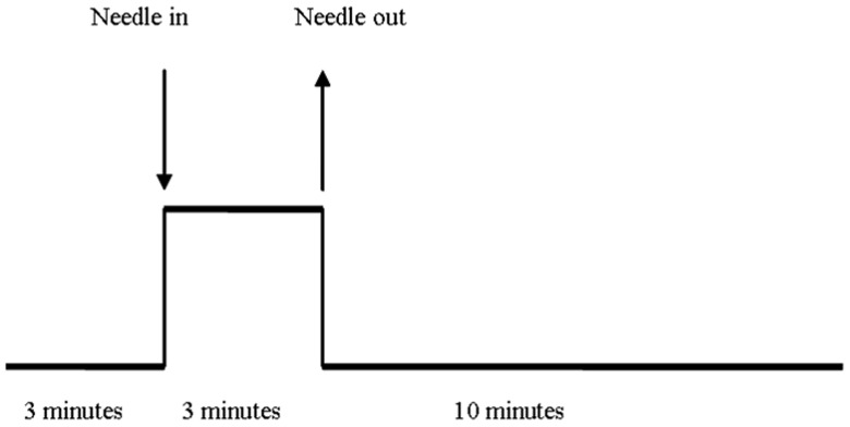
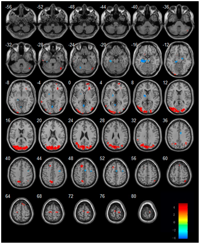
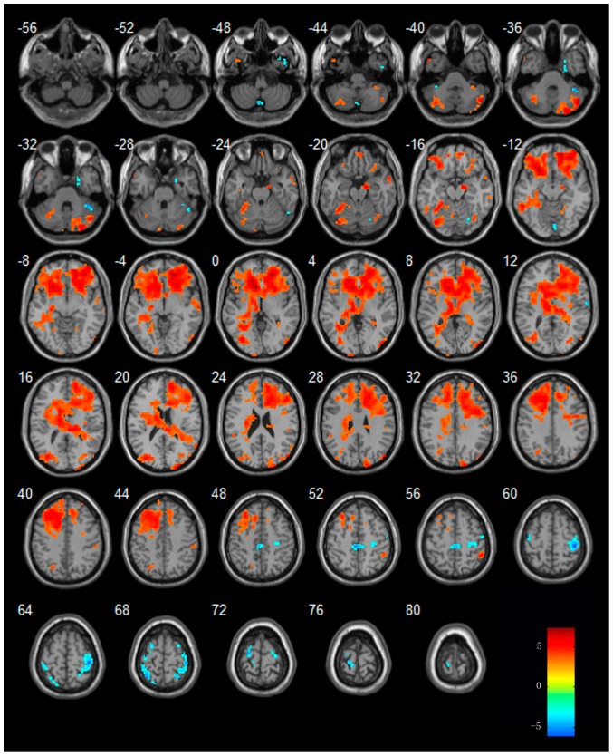
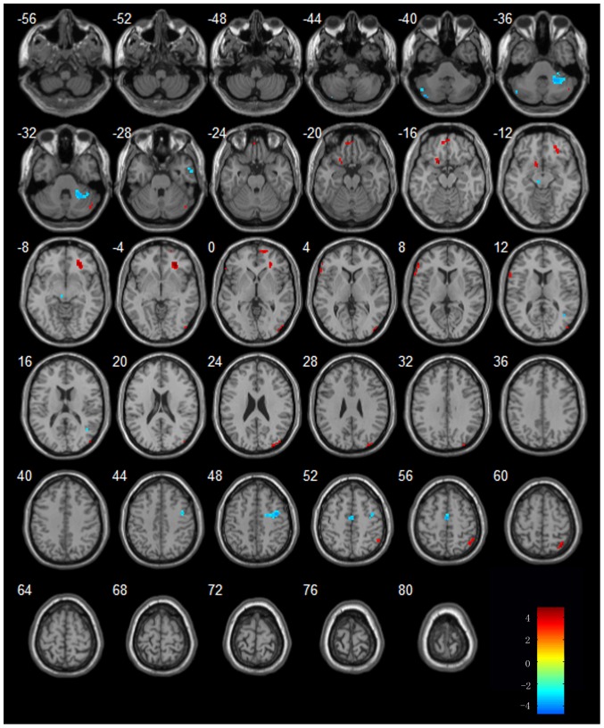
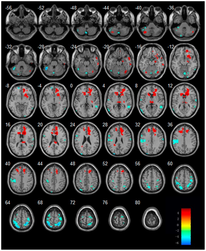
We aim to clarify the mechanisms of acupuncture in treating mild cognitive impairment (MCI) and Alzheimer disease (AD) by using functional magnetic resonance imaging (fMRI). Thirty-six right-handed subjects (8 MCI patients, 14 AD patients, and 14 healthy elders) participated in this study. Clinical and neuropsychological examinations were performed on all the subjects. MRI data acquisition was performed on a SIEMENS verio 3-Tesla scanner. The fMRI study used a single block experimental design. We first acquired the baseline resting state data in the initial 3 minutes; we then acquired the fMRI data during the procession of acupuncture stimulation on the acupoints of Tai chong and Hegu for the following 3 minutes. Last, we acquired fMRI data for another 10 minutes after the needle was withdrawn. The preprocessing and data analysis were performed using the statistical parametric mapping (SPM8) software. Then the two-sample t-tests were performed between each two groups of different states. We found that during the resting state, brain activities in AD and MCI patients were different from those of control subjects. During the acupuncture and the second resting state after acupuncture, when comparing to resting state, there are several regions showing increased or decreased activities in MCI, AD subjects compared to normal subjects. Most of the regions were involved in the temporal lobe and the frontal lobe, which were closely related to the memory and cognition. In conclusion, we investigated the effect of acupuncture in AD and MCI patients by combing fMRI and traditional acupuncture. Our fMRI study confirmed that acupuncture at Tai chong (Liv3) and He gu (LI4) can activate certain cognitive-related regions in AD and MCI patients.
我们旨在通过功能性磁共振成像(fMRI)来阐明针刺治疗轻度认知障碍(MCI)和阿尔茨海默病(AD)的机制。36 名右利手受试者(8 名 MCI 患者、14 名 AD 患者和 14 名健康老年人)参与了这项研究。所有受试者均进行了临床和神经心理学检查。MRI 数据采集在 SIEMENS verio 3-Tesla 扫描仪上进行。fMRI 研究采用单次块实验设计。我们首先在最初的 3 分钟内采集静息状态数据;然后,在针刺太冲和合谷穴位进行针刺刺激的过程中采集 fMRI 数据,持续 3 分钟。最后,在拔针后再采集 fMRI 数据 10 分钟。预处理和数据分析使用统计参数映射(SPM8)软件进行。然后,我们在每个两个不同状态的组之间进行了两样本 t 检验。我们发现,在静息状态下,AD 和 MCI 患者的大脑活动与对照组不同。在针刺和针刺后第二次静息状态期间,与静息状态相比,MCI 和 AD 患者的某些区域的活动增加或减少。大多数区域涉及颞叶和额叶,与记忆和认知密切相关。总之,我们通过结合 fMRI 和传统针刺研究了针刺对 AD 和 MCI 患者的影响。我们的 fMRI 研究证实,针刺太冲(Liv3)和合谷(LI4)可以激活 AD 和 MCI 患者的某些与认知相关的区域。