Department of Pathology, Memorial Sloan-Kettering Cancer Center, New York, NY, USA.
Mod Pathol. 2013 Jan;26(1):131-8. doi: 10.1038/modpathol.2012.138. Epub 2012 Aug 24.
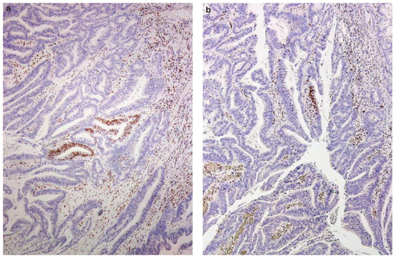
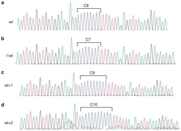
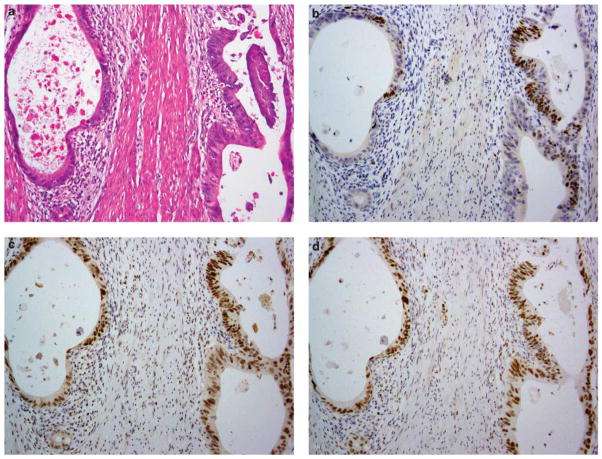
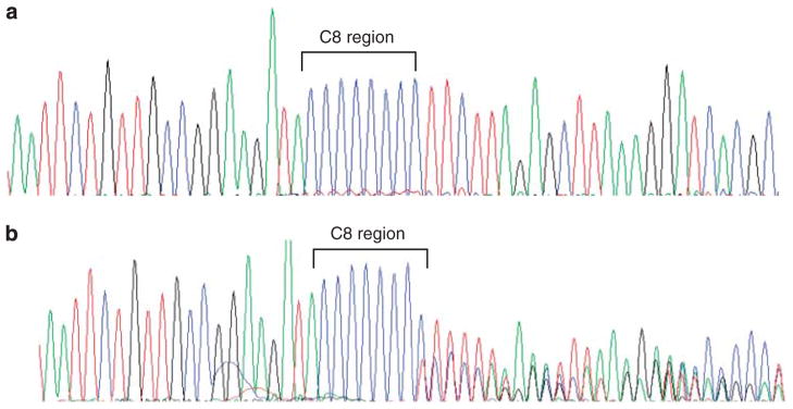
Immunohistochemical staining for DNA mismatch repair proteins may be affected by various biological and technical factors. Staining variations that could potentially lead to erroneous interpretations have been recognized. A recently recognized staining variation is the significant reduction of staining for MSH6 in some colorectal carcinomas. The frequency and specific characteristics of this aberrant MSH6 staining pattern, however, have not been well analyzed. In this study of 420 colorectal carcinoma samples obtained from patients fulfilling the Revised Bethesda Guidelines, we detected 9 tumors (2%) showing extremely limited staining for MSH6 with positive staining present in <5% of the tumor cells. Our analyses showed that these tumors belonged to two distinct categories: (1) MLH1 and/or PMS2 protein-deficient carcinomas (n=5, including 1 with a pathogenic mutation in PMS2); and (2) MLH1, PMS2 and MSH2 normal but with chemotherapy or chemoradiation therapy before surgery (n=4). To test our hypothesis that somatic mutation in the coding region microsatellite of the MSH6 gene might be a potential underlying mechanism for such limited MSH6 staining, we evaluated frameshift mutation in a (C)(8) tract in exon 5 of the MSH6 gene in seven tumors that had sufficient DNA for analysis, and detected mutation in four; all four tumors belonged to the MLH1/PMS2-deficient group. In conclusion, our data outline the main scenarios where significant reduction of MSH6 staining is more likely to occur in colorectal carcinoma, and suggest that somatic mutations of the coding region microsatellites of the MSH6 gene is an underlying mechanism for this staining phenomenon in MLH1/PMS2-deficient carcinomas.
免疫组织化学染色的错配修复蛋白可能会受到各种生物和技术因素的影响。已经认识到可能导致错误解释的染色变化。最近认识到的染色变化是在一些结直肠癌中 MSH6 染色显著减少。然而,这种异常 MSH6 染色模式的频率和具体特征尚未得到很好的分析。在这项对符合修订后的贝塞斯达指南的 420 例结直肠癌患者的研究中,我们检测到 9 例(2%)肿瘤 MSH6 染色非常有限,阳性染色存在于<5%的肿瘤细胞中。我们的分析表明,这些肿瘤属于两种不同的类别:(1)MLH1 和/或 PMS2 蛋白缺陷型癌(n=5,包括 1 例 PMS2 中存在致病性突变);和(2)MLH1、PMS2 和 MSH2 正常,但在手术前接受化疗或放化疗(n=4)。为了验证我们的假设,即 MSH6 基因编码区微卫星中的体细胞突变可能是这种有限 MSH6 染色的潜在机制,我们在 7 例有足够 DNA 进行分析的肿瘤中评估了 MSH6 基因外显子 5 中(C)(8) 重复的移码突变,并在 4 例中检测到突变;所有 4 例肿瘤均属于 MLH1/PMS2 缺陷型组。总之,我们的数据概述了 MSH6 染色显著减少更可能发生在结直肠癌中的主要情况,并表明 MSH6 基因编码区微卫星的体细胞突变是 MLH1/PMS2 缺陷型癌中这种染色现象的潜在机制。