• 文献检索
  • 文档翻译
  • 深度研究
  • 学术资讯
  • Suppr Zotero 插件Zotero 插件
  • 邀请有礼
  • 套餐&价格
  • 历史记录
应用&插件
Suppr Zotero 插件Zotero 插件浏览器插件Mac 客户端Windows 客户端微信小程序
定价
高级版会员购买积分包购买API积分包
服务
文献检索文档翻译深度研究API 文档MCP 服务
关于我们
关于 Suppr公司介绍联系我们用户协议隐私条款
关注我们

Suppr 超能文献

核心技术专利:CN118964589B侵权必究
粤ICP备2023148730 号-1Suppr @ 2026

文献检索

告别复杂PubMed语法,用中文像聊天一样搜索,搜遍4000万医学文献。AI智能推荐,让科研检索更轻松。

立即免费搜索

文件翻译

保留排版,准确专业,支持PDF/Word/PPT等文件格式,支持 12+语言互译。

免费翻译文档

深度研究

AI帮你快速写综述,25分钟生成高质量综述,智能提取关键信息,辅助科研写作。

立即免费体验

心率变异性的心脏内起源、起搏电流及其与危重病的可能关联。

Intracardiac origin of heart rate variability, pacemaker funny current and their possible association with critical illness.

作者信息

Papaioannou Vasilios E, Verkerk Arie O, Amin Ahmed S, de Bakker Jaques M T

机构信息

Democritus University of Thrace, Alexandroupolis University Hospital, Intensive Care Unit, Alexandroupolis Medical School, Dragana 68100, Greece.

出版信息

Curr Cardiol Rev. 2013 Feb 1;9(1):82-96. doi: 10.2174/157340313805076359.

DOI:10.2174/157340313805076359
PMID:22920474
原文链接:https://pmc.ncbi.nlm.nih.gov/articles/PMC3584310/
Abstract

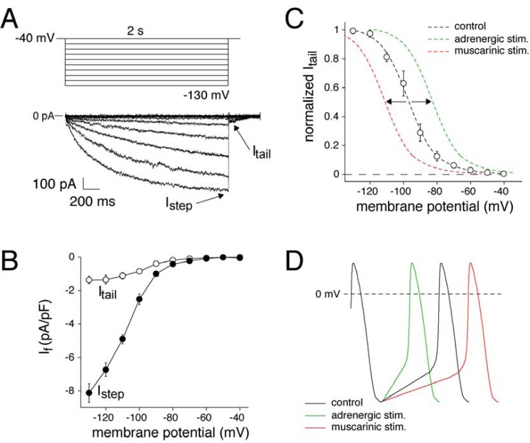
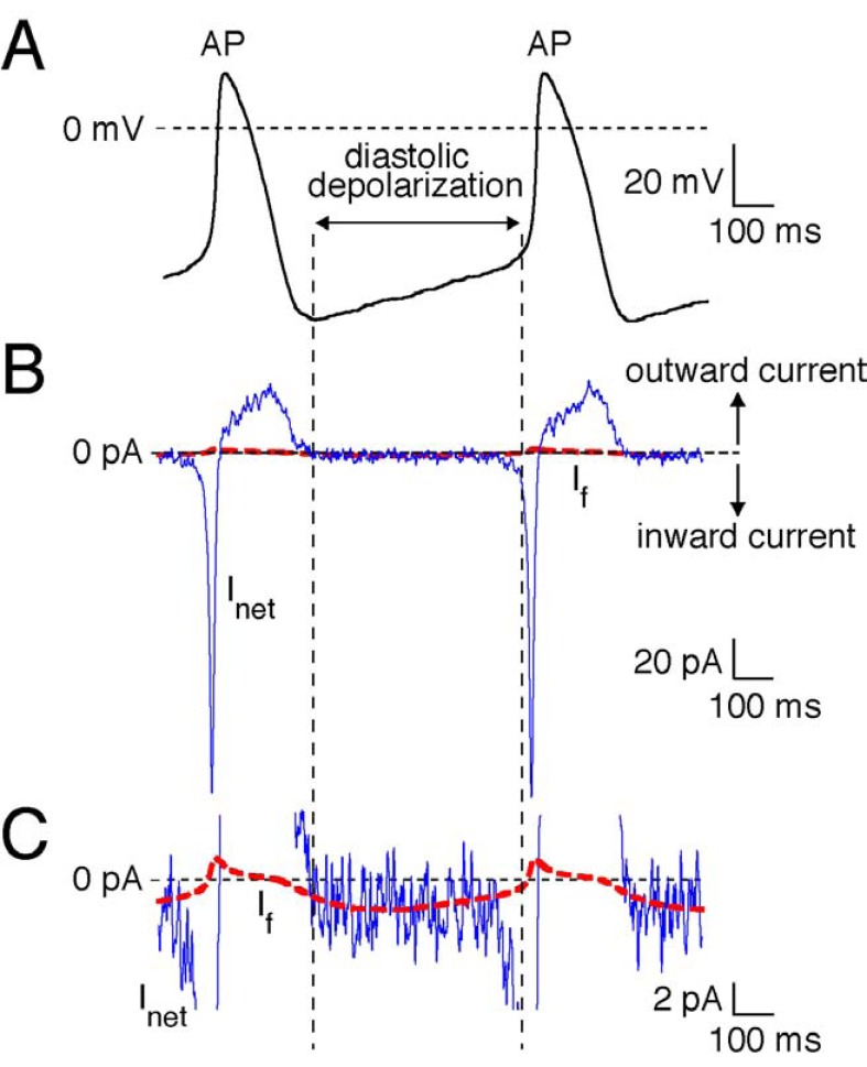
Heart rate variability (HRV) is an indirect estimator of autonomic modulation of heart rate and is considered a risk marker in critical illness, particularly in heart failure and severe sepsis. A reduced HRV has been found in critically ill patients and has been associated with neuro-autonomic uncoupling or decreased baroreflex sensitivity. However, results from human and animal experimental studies indicate that intracardiac mechanisms might also be responsible for interbeat fluctuations. These studies have demonstrated that different membrane channel proteins and especially the so-called 'funny' current (If), an hyperpolarization-activated, inward current that drives diastolic depolarization resulting in spontaneous activity in cardiac pacemaker cells, are altered during critical illness. Furthermore, membrane channels kinetics seem to have significant impact upon HRV, whose early decrease might reflect a cellular metabolic stress. In this review article we present research findings regarding intracardiac origin of HRV, at the cellular level and in both isolated sinoatrial node and whole ex vivo heart preparations. In addition, we will review results from various experimental studies that support the interrelation between If and HRV during endotoxemia. We suggest that reduced HRV during sepsis could also be associated with altered pacemaker cell membrane properties, due to ionic current remodeling.

摘要

心率变异性(HRV)是心率自主调节的一种间接评估指标,被认为是危重病尤其是心力衰竭和严重脓毒症的风险标志物。在危重病患者中发现HRV降低,且与神经自主解耦或压力反射敏感性降低有关。然而,人和动物实验研究的结果表明,心内机制也可能是心跳间期波动的原因。这些研究表明,在危重病期间,不同的膜通道蛋白,特别是所谓的“起搏电流”(If),一种超极化激活的内向电流,它驱动舒张期去极化,导致心脏起搏细胞的自发活动,会发生改变。此外,膜通道动力学似乎对HRV有重大影响,HRV的早期降低可能反映了细胞代谢应激。在这篇综述文章中,我们展示了关于HRV心内起源的研究结果,涉及细胞水平以及分离的窦房结和完整的离体心脏标本。此外,我们将回顾各种实验研究的结果,这些结果支持在内毒素血症期间If与HRV之间的相互关系。我们认为,脓毒症期间HRV降低也可能与起搏细胞膜特性改变有关,这是由于离子电流重塑所致。

https://cdn.ncbi.nlm.nih.gov/pmc/blobs/15ff/3584310/0ca8d150d655/CCR-9-82_F3.jpg
https://cdn.ncbi.nlm.nih.gov/pmc/blobs/15ff/3584310/f0790c456e64/CCR-9-82_F1.jpg
https://cdn.ncbi.nlm.nih.gov/pmc/blobs/15ff/3584310/a4aa6d644ae8/CCR-9-82_F2.jpg
https://cdn.ncbi.nlm.nih.gov/pmc/blobs/15ff/3584310/0ca8d150d655/CCR-9-82_F3.jpg
https://cdn.ncbi.nlm.nih.gov/pmc/blobs/15ff/3584310/f0790c456e64/CCR-9-82_F1.jpg
https://cdn.ncbi.nlm.nih.gov/pmc/blobs/15ff/3584310/a4aa6d644ae8/CCR-9-82_F2.jpg
https://cdn.ncbi.nlm.nih.gov/pmc/blobs/15ff/3584310/0ca8d150d655/CCR-9-82_F3.jpg

相似文献

1
Intracardiac origin of heart rate variability, pacemaker funny current and their possible association with critical illness.心率变异性的心脏内起源、起搏电流及其与危重病的可能关联。
Curr Cardiol Rev. 2013 Feb 1;9(1):82-96. doi: 10.2174/157340313805076359.
2
The funny current: cellular basis for the control of heart rate.滑稽电流:心率控制的细胞基础。
Drugs. 2007;67 Suppl 2:15-24. doi: 10.2165/00003495-200767002-00003.
3
From beat rate variability in induced pluripotent stem cell-derived pacemaker cells to heart rate variability in human subjects.从诱导多能干细胞衍生的起搏细胞中的心率变异性到人类受试者的心率变异性。
Heart Rhythm. 2014 Oct;11(10):1808-1818. doi: 10.1016/j.hrthm.2014.05.037. Epub 2014 Jun 2.
4
Single cells isolated from human sinoatrial node: action potentials and numerical reconstruction of pacemaker current.从人窦房结分离的单细胞:动作电位与起搏电流的数值重建
Annu Int Conf IEEE Eng Med Biol Soc. 2007;2007:904-7. doi: 10.1109/IEMBS.2007.4352437.
5
Depressed pacemaker activity of sinoatrial node myocytes contributes to the age-dependent decline in maximum heart rate.窦房结心肌细胞抑郁的起搏活动导致最大心率随年龄的增长而下降。
Proc Natl Acad Sci U S A. 2013 Oct 29;110(44):18011-6. doi: 10.1073/pnas.1308477110. Epub 2013 Oct 15.
6
Cardiac pacemaker I(f) current and its inhibition by heart rate-reducing agents.心脏起搏器I(f)电流及其被心率降低剂抑制的情况。
Curr Med Res Opin. 2005 Jul;21(7):1115-22. doi: 10.1185/030079905x50543.
7
Pacemaker activity of the human sinoatrial node: role of the hyperpolarization-activated current, I(f).人类窦房结的起搏活动:超极化激活电流I(f)的作用。
Int J Cardiol. 2009 Mar 6;132(3):318-36. doi: 10.1016/j.ijcard.2008.12.196. Epub 2009 Jan 31.
8
The role of the funny current in pacemaker activity.有趣电流在起搏器活动中的作用。
Circ Res. 2010 Feb 19;106(3):434-46. doi: 10.1161/CIRCRESAHA.109.208041.
9
Beating irregularity of single pacemaker cells isolated from the rabbit sinoatrial node.从兔窦房结分离出的单个起搏细胞的搏动不规则性。
Biophys J. 1993 Dec;65(6):2601-13. doi: 10.1016/S0006-3495(93)81289-X.
10
Altered heart rate variability in angiotensin II-mediated hypertension is associated with impaired autonomic nervous system signaling and intrinsic sinoatrial node dysfunction.血管紧张素 II 介导的高血压中心率变异性改变与自主神经系统信号传递受损和固有窦房结功能障碍有关。
Heart Rhythm. 2020 Aug;17(8):1360-1370. doi: 10.1016/j.hrthm.2020.03.014. Epub 2020 Mar 27.

引用本文的文献

1
Slow nocturnal body cooling during sleep increases interbeat intervals and is tightly coupled to high-frequency heart rate variability in healthy men.睡眠期间夜间身体缓慢降温会增加心跳间期,并且与健康男性的高频心率变异性紧密相关。
Physiol Rep. 2025 Aug;13(15):e70478. doi: 10.14814/phy2.70478.
2
Publication guidelines for human heart rate and heart rate variability studies in psychophysiology-Part 1: Physiological underpinnings and foundations of measurement.人体心率和心率变异性研究的出版指南——第 1 部分:生理基础和测量基础。
Psychophysiology. 2024 Sep;61(9):e14604. doi: 10.1111/psyp.14604. Epub 2024 Jun 14.
3

本文引用的文献

1
Reducing elevated heart rate in patients with multiple organ dysfunction syndrome by the I (f) (funny channel current) inhibitor ivabradine : MODI (f)Y trial.应用 I(f)(有趣通道电流)抑制剂伊伐布雷定降低多器官功能障碍综合征患者的心率:MODI(f)Y 试验。
Clin Res Cardiol. 2011 Oct;100(10):915-23. doi: 10.1007/s00392-011-0323-2. Epub 2011 Jun 3.
2
Differential reduction of HCN channel activity by various types of lipopolysaccharide.各种类型的脂多糖对 HCN 通道活性的差异降低作用。
J Mol Cell Cardiol. 2011 Aug;51(2):226-35. doi: 10.1016/j.yjmcc.2011.05.004. Epub 2011 May 14.
3
Heart rate as a risk factor in chronic heart failure (SHIFT): the association between heart rate and outcomes in a randomised placebo-controlled trial.
Somatodendritic organization of pacemaker activity in midbrain dopamine neurons.
中脑多巴胺能神经元中起搏器活动的体树突组织
Korean J Physiol Pharmacol. 2024 Mar 1;28(2):165-181. doi: 10.4196/kjpp.2024.28.2.165.
4
Cardiovascular autonomic dysfunction in "Long COVID": pathophysiology, heart rate variability, and inflammatory markers.“长新冠”中的心血管自主神经功能障碍:病理生理学、心率变异性和炎症标志物
Front Cardiovasc Med. 2023 Sep 1;10:1256512. doi: 10.3389/fcvm.2023.1256512. eCollection 2023.
5
A Narrative Review of Commercial Platforms Offering Tracking of Heart Rate Variability in Corporate Employees to Detect and Manage Stress.关于为企业员工提供心率变异性追踪以检测和管理压力的商业平台的叙述性综述。
J Cardiovasc Dev Dis. 2023 Mar 27;10(4):141. doi: 10.3390/jcdd10040141.
6
Decreased Heart Rate Variability in COVID-19.新型冠状病毒肺炎患者心率变异性降低
Intensive Care Res. 2023;3(1):87-91. doi: 10.1007/s44231-022-00024-1. Epub 2022 Dec 1.
7
Evaluation of non-linear heart rate variability using multi-scale multi-fractal detrended fluctuation analysis in mice: Roles of the autonomic nervous system and sinoatrial node.使用多尺度多重分形去趋势波动分析评估小鼠的非线性心率变异性:自主神经系统和窦房结的作用。
Front Physiol. 2022 Sep 27;13:970393. doi: 10.3389/fphys.2022.970393. eCollection 2022.
8
Increased variability of fetal heart rate during labour: a review of preclinical and clinical studies.分娩过程中胎儿心率变异性增加:临床前和临床研究综述。
BJOG. 2022 Nov;129(12):2070-2081. doi: 10.1111/1471-0528.17234. Epub 2022 Jun 5.
9
Fractal correlation properties of heart rate variability as a biomarker of endurance exercise fatigue in ultramarathon runners.心率变异性的分形相关性特征可作为超长马拉松运动员耐力运动性疲劳的生物标志物。
Physiol Rep. 2021 Jul;9(14):e14956. doi: 10.14814/phy2.14956.
10
Natriuretic peptide receptor B maintains heart rate and sinoatrial node function via cyclic GMP-mediated signalling.利钠肽受体 B 通过环鸟苷酸介导的信号转导维持心率和窦房结功能。
Cardiovasc Res. 2022 Jun 29;118(8):1917-1931. doi: 10.1093/cvr/cvab245.
心率作为慢性心力衰竭的危险因素(SHIFT):一项随机安慰剂对照试验中心率与结局的关系。
Lancet. 2010 Sep 11;376(9744):886-94. doi: 10.1016/S0140-6736(10)61259-7.
4
Lipopolysaccharides directly decrease Ca2+ oscillations and the hyperpolarization-activated nonselective cation current If in immortalized HL-1 cardiomyocytes.脂多糖直接降低 HL-1 心肌细胞中的钙离子振荡和超极化激活非选择性阳离子电流 If。
Am J Physiol Cell Physiol. 2010 Sep;299(3):C665-71. doi: 10.1152/ajpcell.00129.2010. Epub 2010 Jun 23.
5
Fractals for physicians.医生的分形学。
Paediatr Respir Rev. 2010 Jun;11(2):123-31. doi: 10.1016/j.prrv.2010.02.001. Epub 2010 Mar 4.
6
Endotoxin depresses heart rate variability in mice: cytokine and steroid effects.内毒素降低小鼠心率变异性:细胞因子和类固醇的作用。
Am J Physiol Regul Integr Comp Physiol. 2009 Oct;297(4):R1019-27. doi: 10.1152/ajpregu.00132.2009. Epub 2009 Aug 5.
7
Generality of a power-law long-term correlation in beat timings of single cardiac cells.单个心肌细胞搏动时间中幂律长期相关性的普遍性。
Biochem Biophys Res Commun. 2009 Sep 11;387(1):19-24. doi: 10.1016/j.bbrc.2009.05.143. Epub 2009 Jun 6.
8
Impaired regulation of cardiac function in sepsis, SIRS, and MODS.脓毒症、全身炎症反应综合征和多器官功能障碍综合征中心脏功能调节受损。
Can J Physiol Pharmacol. 2009 Apr;87(4):266-74. doi: 10.1139/Y09-012.
9
Associated changes in HCN2 and HCN4 transcripts and I(f) pacemaker current in myocytes.心肌细胞中HCN2和HCN4转录本及I(f)起搏电流的相关变化。
Biochim Biophys Acta. 2009 May;1788(5):1138-47. doi: 10.1016/j.bbamem.2009.02.011. Epub 2009 Feb 21.
10
Pacemaker activity of the human sinoatrial node: role of the hyperpolarization-activated current, I(f).人类窦房结的起搏活动:超极化激活电流I(f)的作用。
Int J Cardiol. 2009 Mar 6;132(3):318-36. doi: 10.1016/j.ijcard.2008.12.196. Epub 2009 Jan 31.