Department of Orthopedic Surgery, Asan Medical Center, University of Ulsan College of Medicine, Seoul, Korea.
Clin Orthop Surg. 2012 Sep;4(3):209-15. doi: 10.4055/cios.2012.4.3.209. Epub 2012 Aug 14.
We conducted this radiographic study in the elderly population with proximal humeral fracture aiming to evaluate 1) the serial changes of neck-shaft angle after locking plate fixation and 2) find relationship between change in neck shaft angle and various factors such as age, fracture pattern, severity of osteoporosis, medial support and initial reduction angle.
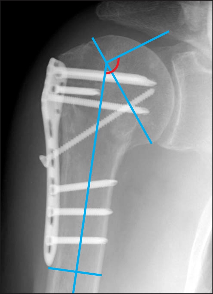
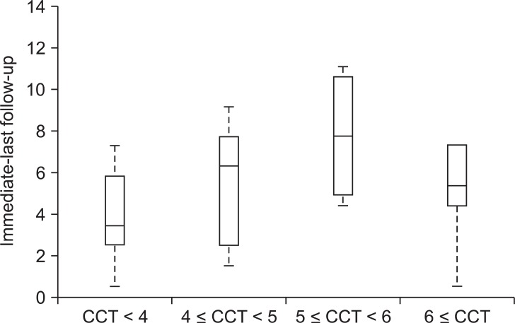
Twenty-five patients who underwent surgical treatment for proximal humeral fracture with locking plate between September 2008 and August 2010 are included. True anteroposterior and axillary lateral radiographs were made postoperatively and at each follow-up visit. Measurement of neck shaft angle was done at immediate postoperative, 3 months postoperative and a final follow-up (average, 11 months; range, 8 to 17 months). Severity of osteoporosis was assessed using cortical thickness suggested by Tingart et al.
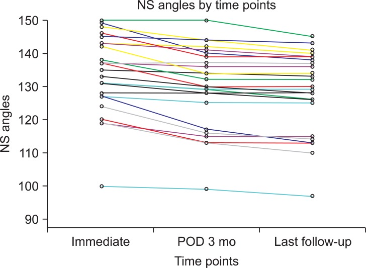
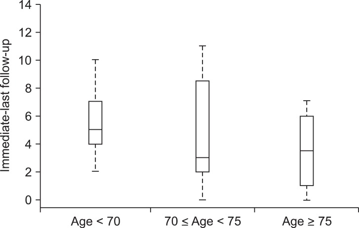
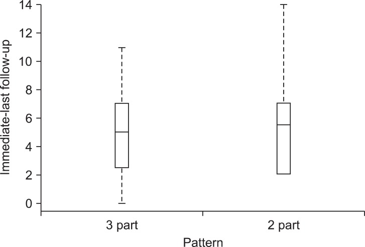
The mean neck shaft angles were 133.6° (range, 100° to 116°) at immediate postoperative, 129.8° (range, 99° to 150°) at 3 months postoperative and 128.4° (range, 97° to 145°) at final follow-up. The mean loss in the neck-shaft angle in the first 3 months was 3.8° as compared to 1.3° in the period between 3 months and final follow-up. This was statistically significant (p = 0.002), indicating that most of the fall in neck shaft angle occurs in the first three months after surgery. Relationship between neck shaft angle change and age (p = 0.29), fracture pattern (p = 0.41), cortical thickness (p = 0.21), medial support (p = 0.63) and initial reduction accuracy (p = 0.65) are not statistically significant.
The proximal humerus locking plate maintains reliable radiographic results even in the elderly population with proximal humerus fracture.
我们对老年肱骨近端骨折患者进行了这项放射学研究,旨在评估 1)锁定钢板固定后颈干角的连续变化,2)发现颈干角变化与年龄、骨折类型、骨质疏松严重程度、内侧支撑和初始复位角度等各种因素之间的关系。
2008 年 9 月至 2010 年 8 月期间,我们对 25 例接受锁定钢板治疗的肱骨近端骨折患者进行了手术治疗。术后和每次随访时均拍摄正位和腋位 X 线片。术后即刻、术后 3 个月和最终随访(平均 11 个月;范围 8 至 17 个月)时测量颈干角。骨质疏松严重程度采用 Tingart 等提出的皮质厚度评估。
术后即刻颈干角平均为 133.6°(范围 100°至 116°),术后 3 个月为 129.8°(范围 99°至 150°),最终随访时为 128.4°(范围 97°至 145°)。与术后 3 个月至最终随访期间的 1.3°相比,术后 3 个月内颈干角平均丢失 3.8°。这具有统计学意义(p = 0.002),表明颈干角的大部分下降发生在术后 3 个月内。颈干角变化与年龄(p = 0.29)、骨折类型(p = 0.41)、皮质厚度(p = 0.21)、内侧支撑(p = 0.63)和初始复位准确性(p = 0.65)之间无统计学意义。
即使在老年肱骨近端骨折患者中,肱骨近端锁定钢板也能保持可靠的影像学结果。