• 文献检索
  • 文档翻译
  • 深度研究
  • 学术资讯
  • Suppr Zotero 插件Zotero 插件
  • 邀请有礼
  • 套餐&价格
  • 历史记录
应用&插件
Suppr Zotero 插件Zotero 插件浏览器插件Mac 客户端Windows 客户端微信小程序
定价
高级版会员购买积分包购买API积分包
服务
文献检索文档翻译深度研究API 文档MCP 服务
关于我们
关于 Suppr公司介绍联系我们用户协议隐私条款
关注我们

Suppr 超能文献

核心技术专利:CN118964589B侵权必究
粤ICP备2023148730 号-1Suppr @ 2026

文献检索

告别复杂PubMed语法,用中文像聊天一样搜索,搜遍4000万医学文献。AI智能推荐,让科研检索更轻松。

立即免费搜索

文件翻译

保留排版,准确专业,支持PDF/Word/PPT等文件格式,支持 12+语言互译。

免费翻译文档

深度研究

AI帮你快速写综述,25分钟生成高质量综述,智能提取关键信息,辅助科研写作。

立即免费体验

非综合征性唇腭裂患者咽气道的三维评估。

Three dimensional assessment of the pharyngeal airway in individuals with non-syndromic cleft lip and palate.

机构信息

University of California Los Angeles, Los Angeles, California, United States of America.

出版信息

PLoS One. 2012;7(8):e43405. doi: 10.1371/journal.pone.0043405. Epub 2012 Aug 29.

DOI:10.1371/journal.pone.0043405
PMID:22952677
原文链接:https://pmc.ncbi.nlm.nih.gov/articles/PMC3430706/
Abstract

INTRODUCTION

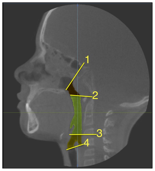
Children with cleft lip and palate (CLP) are known to have airway problems. Previous studies have shown that individuals with CLP have a 30% reduction in nasal airway size compared to non-cleft controls. No reports have been found on cross-sectional area and volume of the pharyngeal airway in clefts. Introduction of Cone-Beam CT (CBCT) and imaging software has facilitated generation of 3D images for assessment of the cross-sectional area and volume of the airway.

OBJECTIVE

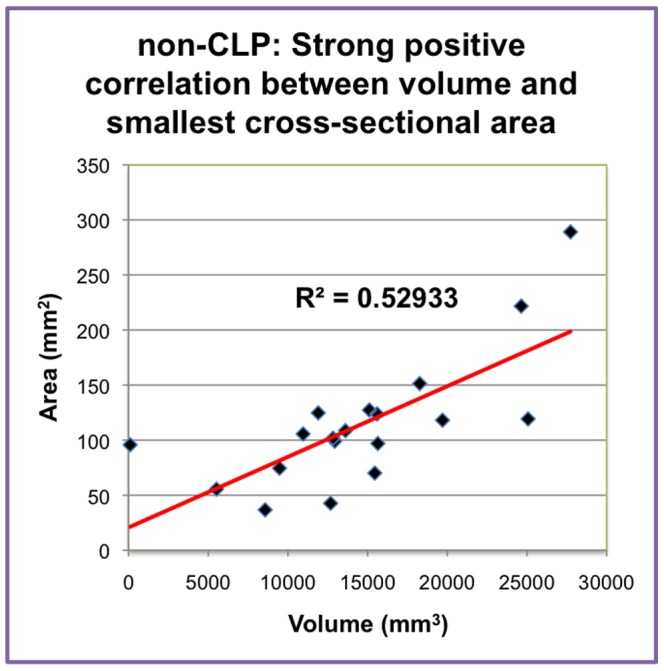
To assess the pharyngeal airway in individuals with CLP using CBCT by measuring volume and smallest cross-sectional areas and compare with 19 age- and sex-matched non-cleft controls.

METHODS

Retrospective study of CBCT data of pre-adolescent individuals (N = 19, Mean age = 10.6, 7 females, 12 males, UCLP = 6, BCLP = 3) from the Center for Craniofacial Anomalies. Volumetric analysis was performed using image segmentation features in CB Works 3.0. Volume and smallest cross-sectional were studied in both groups. Seven measurements were repeated to verify reliability using Pearson correlation coefficient. Volume and cross-sectional area differences were analyzed using paired t-tests.

RESULTS

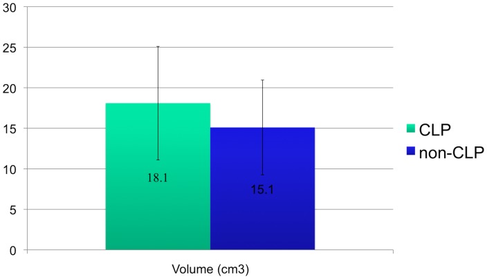
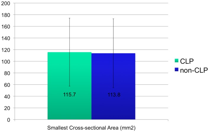
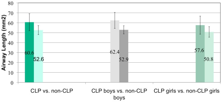
The method was found to be reliable. Individuals with CLP did not exhibit smaller total airway volume and cross sectional area than non-CLP controls.

CONCLUSION

3D imaging using CBCT and CB Works is reliable for assessing airway volume. Previous studies have shown that the nasal airway is restricted in individuals with CLP. In our study, we found that the pharyngeal airway is not compromised in these individuals.

摘要

简介

患有唇腭裂(CLP)的儿童已知存在气道问题。先前的研究表明,与非裂隙对照组相比,CLP 个体的鼻气道尺寸减小了 30%。目前尚无关于裂隙患者咽气道横截面积和体积的报告。锥形束 CT(CBCT)和成像软件的引入促进了气道横截面积和体积的 3D 图像生成,以进行评估。

目的

使用 CBCT 通过测量体积和最小横截面积来评估 CLP 个体的咽气道,并与 19 名年龄和性别匹配的非裂隙对照组进行比较。

方法

对来自颅面畸形中心的青少年前个体(N = 19,平均年龄 10.6,7 名女性,12 名男性,UCLP = 6,BCLP = 3)的 CBCT 数据进行回顾性研究。使用 CB Works 3.0 中的图像分割功能进行容积分析。在两组中都研究了体积和最小横截面积。使用 Pearson 相关系数重复了七次测量以验证可靠性。使用配对 t 检验分析体积和横截面积差异。

结果

该方法被证明是可靠的。CLP 个体的总气道体积和横截面积并不小于非 CLP 对照组。

结论

使用 CBCT 和 CB Works 的 3D 成像对于评估气道体积是可靠的。先前的研究表明,CLP 个体的鼻气道受到限制。在我们的研究中,我们发现这些个体的咽气道没有受到影响。

https://cdn.ncbi.nlm.nih.gov/pmc/blobs/85cb/3430706/e917c61341a5/pone.0043405.g005.jpg
https://cdn.ncbi.nlm.nih.gov/pmc/blobs/85cb/3430706/6c87b6605089/pone.0043405.g001.jpg
https://cdn.ncbi.nlm.nih.gov/pmc/blobs/85cb/3430706/30eba7b28247/pone.0043405.g002.jpg
https://cdn.ncbi.nlm.nih.gov/pmc/blobs/85cb/3430706/feaecee1606b/pone.0043405.g003.jpg
https://cdn.ncbi.nlm.nih.gov/pmc/blobs/85cb/3430706/41991c0b880e/pone.0043405.g004.jpg
https://cdn.ncbi.nlm.nih.gov/pmc/blobs/85cb/3430706/e917c61341a5/pone.0043405.g005.jpg

相似文献

1
Three dimensional assessment of the pharyngeal airway in individuals with non-syndromic cleft lip and palate.非综合征性唇腭裂患者咽气道的三维评估。
PLoS One. 2012;7(8):e43405. doi: 10.1371/journal.pone.0043405. Epub 2012 Aug 29.
2
Cone Beam Computed Tomography Analysis of Oropharyngeal Airway in Preadolescent Nonsyndromic Bilateral and Unilateral Cleft Lip and Palate Patients.青春期前非综合征性双侧和单侧唇腭裂患者口咽气道的锥形束计算机断层扫描分析
Cleft Palate Craniofac J. 2018 Jul;55(6):883-890. doi: 10.1597/15-322. Epub 2018 Feb 22.
3
Volumetric nasal cavity analysis in children with unilateral and bilateral cleft lip and palate.单侧和双侧唇腭裂患儿鼻腔容积分析
Laryngoscope. 2016 Jun;126(6):1475-80. doi: 10.1002/lary.25543. Epub 2015 Aug 12.
4
Three-dimensional assessment of airway volumes in patients with unilateral cleft lip and palate.单侧唇腭裂患者气道容积的三维评估。
Prog Orthod. 2021 Nov 8;22(1):35. doi: 10.1186/s40510-021-00382-4.
5
Comparative Evaluation of the Pharyngeal Airway Space in Unilateral and Bilateral Cleft Lip and Palate Individuals With Noncleft Individuals: A Cone Beam Computed Tomography Study.单侧和双侧唇腭裂个体与非腭裂个体咽气道空间的比较评估:一项锥形束计算机断层扫描研究
Cleft Palate Craniofac J. 2017 Sep;54(5):509-516. doi: 10.1597/16-013. Epub 2016 Jul 21.
6
Three-dimensional assessment of the pharyngeal airway in Japanese preschoolers with orofacial clefts.日本学龄前腭裂儿童咽腔气道的三维评估。
Laryngoscope. 2020 Feb;130(2):533-540. doi: 10.1002/lary.27957. Epub 2019 Apr 12.
7
Assessment of pharyngeal airway volume in adolescent patients affected by bilateral cleft lip and palate using cone beam computed tomography.使用锥形束计算机断层扫描评估双侧唇腭裂青少年患者的咽气道容积
Angle Orthod. 2014 Nov;84(6):995-1001. doi: 10.2319/121913-930.1. Epub 2014 Mar 25.
8
Evaluation of Airway Volume in Cleft Lip and Palate Following Nasoalveolar Molding.鼻牙槽骨塑形术后唇腭裂气道容积的评估
J Craniofac Surg. 2018 Nov;29(8):2143-2147. doi: 10.1097/SCS.0000000000004681.
9
Comparison of Nasopharyngeal Airway Volume in Cleft Lip and Palate Patients With Normal Individuals Using Cone Beam Computed Tomography.应用锥形束 CT 对正常人群与唇腭裂患者的鼻咽腔容积进行比较。
J Craniofac Surg. 2023 May 1;34(3):1111-1114. doi: 10.1097/SCS.0000000000009055. Epub 2022 Oct 10.
10
Tomographic Pharyngeal Dimensions in Individuals With Unilateral Cleft Lip/Palate and Class III Malocclusion Are Reduced When Compared With Controls.与对照组相比,单侧唇腭裂合并III类错牙合患者的咽部断层扫描尺寸减小。
Cleft Palate Craniofac J. 2017 Sep;54(5):502-508. doi: 10.1597/15-124. Epub 2016 May 5.

引用本文的文献

1
Correlation between different boundaries used in upper airway assessment.上气道评估中使用的不同边界之间的相关性。
BMC Oral Health. 2025 Jan 4;25(1):19. doi: 10.1186/s12903-024-05402-3.
2
Three-dimensional assessment of the nasopharyngeal airway in Down syndrome during the mixed dentition period: a case-control study.混合牙列期唐氏综合征患者鼻咽气道的三维评估:一项病例对照研究。
Angle Orthod. 2025 Jan 1;95(1):78-85. doi: 10.2319/022424-142.1.
3
The usefulness of cone beam computed tomography according to age in cleft lip and palate.

本文引用的文献

1
Three-dimensional assessment of impacted canines and root resorption using cone beam computed tomography.应用锥形束 CT 对埋伏阻生牙进行三维评估和牙根吸收的研究。
Oral Surg Oral Med Oral Pathol Oral Radiol. 2012 Feb;113(2):260-7. doi: 10.1016/j.tripleo.2011.03.035. Epub 2011 Jun 25.
2
Airway and respiratory complications in children undergoing cleft lip and palate repair.唇腭裂修复术患儿的气道及呼吸并发症
Ghana Med J. 2010 Mar;44(1):16-20. doi: 10.4314/gmj.v44i1.68851.
3
Three-dimensional analysis of pharyngeal airway in preadolescent children with different anteroposterior skeletal patterns.
根据年龄评估腭裂患者锥形束 CT 的有用性。
J Med Life. 2022 Sep;15(9):1136-1142. doi: 10.25122/jml-2022-0209.
4
Three-dimensional assessment of airway volumes in patients with unilateral cleft lip and palate.单侧唇腭裂患者气道容积的三维评估。
Prog Orthod. 2021 Nov 8;22(1):35. doi: 10.1186/s40510-021-00382-4.
5
Three-dimensional analysis of the velopharyngeal region in patients with cleft palate and healthy individuals.腭裂患者与正常个体的腭咽区的三维分析。
Surg Radiol Anat. 2020 Sep;42(9):1033-1042. doi: 10.1007/s00276-020-02526-3. Epub 2020 Jul 8.
6
Effect of maxillary expansion and protraction on the oropharyngeal airway in individuals with non-syndromic cleft palate with or without cleft lip.上颌扩张和牵引对上颌裂和非上颌裂腭裂患者口咽气道的影响。
PLoS One. 2019 Jul 30;14(7):e0213328. doi: 10.1371/journal.pone.0213328. eCollection 2019.
7
Volumetric reconstruction and determination of minimum crosssectional area of the pharynx in patients with cleft lip and palate: comparison between two different softwares.唇腭裂患者咽部的容积重建及最小横截面积测定:两种不同软件的比较
J Appl Oral Sci. 2018 Oct 4;26:e20170282. doi: 10.1590/1678-7757-2017-0282.
8
Comparison of the Pharyngeal Airway Volume between Non-Syndromic Unilateral Cleft Palate and Normal Individuals Using Cone Beam Computed Tomography.使用锥形束计算机断层扫描比较非综合征性单侧腭裂患者与正常人的咽气道容积
J Dent (Shiraz). 2016 Sep;17(3 Suppl):268-275.
9
Three-Dimensional Evaluation of the Upper Airway Morphological Changes in Growing Patients with Skeletal Class III Malocclusion Treated by Protraction Headgear and Rapid Palatal Expansion: A Comparative Research.用前方牵引器和快速腭开展治疗的生长发育期骨性III类错牙合患者上气道形态变化的三维评估:一项对比研究
PLoS One. 2015 Aug 7;10(8):e0135273. doi: 10.1371/journal.pone.0135273. eCollection 2015.
10
Three-dimensional imaging methods for quantitative analysis of facial soft tissues and skeletal morphology in patients with orofacial clefts: a systematic review.口腔颌面裂患者面部软组织和骨骼形态定量分析的三维成像方法:一项系统评价
PLoS One. 2014 Apr 7;9(4):e93442. doi: 10.1371/journal.pone.0093442. eCollection 2014.
不同前后向骨骼模式的青春期前儿童咽气道的三维分析。
Am J Orthod Dentofacial Orthop. 2010 Mar;137(3):306.e1-11; discussion 306-7. doi: 10.1016/j.ajodo.2009.10.025.
4
Three-dimensional assessment of the eruption path of the canine in individuals with bone-grafted alveolar clefts using cone beam computed tomography.使用锥形束计算机断层扫描对骨移植牙槽裂患者犬齿萌出路径进行三维评估。
Cleft Palate Craniofac J. 2010 Sep;47(5):507-12. doi: 10.1597/08-171.
5
Automated 3-dimensional airway analysis from cone-beam computed tomography data.基于锥形束计算机断层扫描数据的气道自动三维分析
J Oral Maxillofac Surg. 2010 Mar;68(3):696-701. doi: 10.1016/j.joms.2009.07.040.
6
Comparison of cone-beam CT parameters and sleep questionnaires in sleep apnea patients and control subjects.睡眠呼吸暂停患者与对照受试者的锥束CT参数与睡眠问卷比较。
Oral Surg Oral Med Oral Pathol Oral Radiol Endod. 2010 Feb;109(2):285-93. doi: 10.1016/j.tripleo.2009.09.033.
7
Volumetric assessment of secondary alveolar bone grafting using cone beam computed tomography.使用锥形束计算机断层扫描对二期牙槽骨植骨进行容积评估。
Cleft Palate Craniofac J. 2009 Sep;46(5):503-11. doi: 10.1597/08-153.1. Epub 2009 Feb 27.
8
Oropharyngeal airway in children with Class III malocclusion evaluated by cone-beam computed tomography.通过锥形束计算机断层扫描评估Ⅲ类错牙合儿童的口咽气道。
Am J Orthod Dentofacial Orthop. 2009 Sep;136(3):318.e1-9; discussion 318-9. doi: 10.1016/j.ajodo.2009.02.017.
9
Comparison of airway space with conventional lateral headfilms and 3-dimensional reconstruction from cone-beam computed tomography.气道空间与传统头颅侧位片及锥形束计算机断层扫描三维重建的比较
Am J Orthod Dentofacial Orthop. 2009 Apr;135(4):468-79. doi: 10.1016/j.ajodo.2007.04.043.
10
Age-related changes of the upper airway assessed by 3-dimensional computed tomography.通过三维计算机断层扫描评估上气道的年龄相关变化。
J Craniofac Surg. 2009 Mar;20 Suppl 1:657-63. doi: 10.1097/SCS.0b013e318193d521.