Max Planck Institute for Human Cognitive and Brain Sciences, Leipzig, Germany.
PLoS One. 2012;7(8):e44195. doi: 10.1371/journal.pone.0044195. Epub 2012 Aug 29.
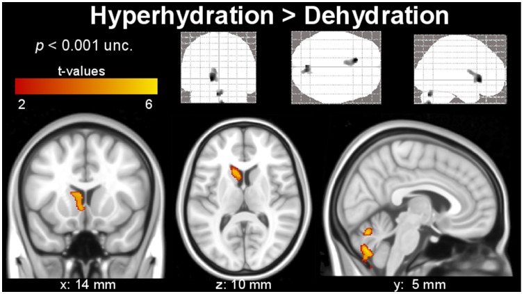
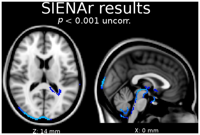
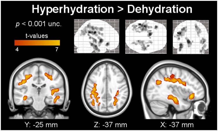
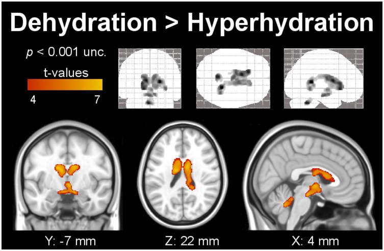
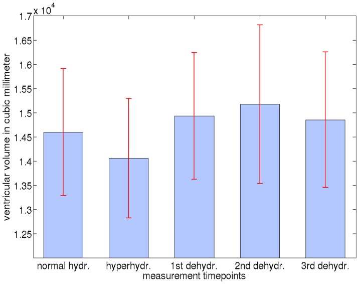
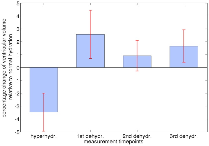
Dehydration can affect the volume of brain structures, which might imply a confound in volumetric and morphometric studies of normal or diseased brain. Six young, healthy volunteers were repeatedly investigated using three-dimensional T(1)-weighted magnetic resonance imaging during states of normal hydration, hyperhydration, and dehydration to assess volume changes in gray matter (GM), white matter (WM), and cerebrospinal fluid (CSF). The datasets were analyzed using voxel-based morphometry (VBM), a widely used voxel-wise statistical analysis tool, FreeSurfer, a fully automated volumetric segmentation measure, and SIENAr a longitudinal brain-change detection algorithm. A significant decrease of GM and WM volume associated with dehydration was found in various brain regions, most prominently, in temporal and sub-gyral parietal areas, in the left inferior orbito-frontal region, and in the extra-nuclear region. Moreover, we found consistent increases in CSF, that is, an expansion of the ventricular system affecting both lateral ventricles, the third, and the fourth ventricle. Similar degrees of shrinkage in WM volume and increase of the ventricular system have been reported in studies of mild cognitive impairment or Alzheimer's disease during disease progression. Based on these findings, a potential confound in GM and WM or ventricular volume studies due to the subjects' hydration state cannot be excluded and should be appropriately addressed in morphometric studies of the brain.
脱水会影响脑结构的体积,这可能会对正常或患病大脑的体积和形态计量研究造成混淆。为了评估灰质 (GM)、白质 (WM) 和脑脊液 (CSF) 的体积变化,六名年轻、健康的志愿者在正常水合、高水合和脱水状态下使用三维 T1 加权磁共振成像进行了反复研究。使用基于体素的形态计量学 (VBM)、一种广泛使用的体素统计分析工具、FreeSurfer(一种全自动体积分割测量)和 SIENAr(一种纵向脑变化检测算法)对数据集进行了分析。在各种脑区发现了与脱水相关的 GM 和 WM 体积显著减少,最明显的是颞叶和皮质下顶叶区域、左侧下眶额区域和核外区域。此外,我们还发现 CSF 持续增加,即脑室系统扩张,影响两侧脑室、第三脑室和第四脑室。在轻度认知障碍或阿尔茨海默病的疾病进展研究中,也报道了 WM 体积相似程度的收缩和脑室系统的增加。基于这些发现,由于受试者的水合状态,GM 和 WM 或脑室体积研究中可能存在混淆因素,在大脑的形态计量研究中应适当解决。