Department of Radiation Oncology, The Cancer Institute of New Jersey, 195 Little Albany Street, New Brunswick, NJ 08903, USA.
J Appl Clin Med Phys. 2012 Sep 6;13(5):3976. doi: 10.1120/jacmp.v13i5.3976.
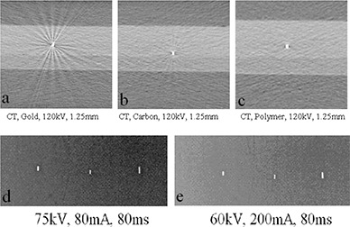
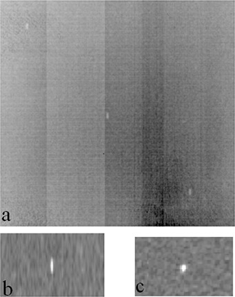
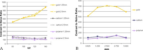
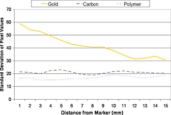
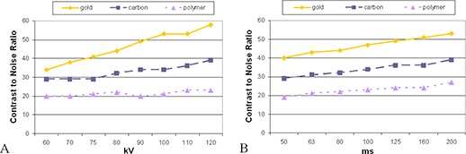
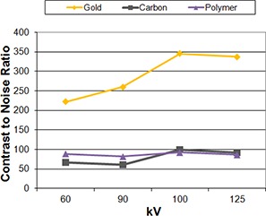
The purpose of this study was to evaluate the visibility and artifact created by gold, carbon, and polymer fiducial markers in a simple phantom across computed tomography (CT), kilovoltage (kV), and megavoltage (MV) linear accelerator imaging and MV tomotherapy imaging. Three types of fiducial markers (gold, carbon, and polymer) were investigated for their visibility and artifacts in images acquired with various modalities and with different imaging parameters (kV, mAs, slice thickness). The imaging modalities include kV CT, 2D linac-based kilovoltage and megavoltage X-ray imaging systems, kV cone-beam CT, and normal and fine tomotherapy imaging. The images were acquired on a phantom constructed using Superflab bolus in which markers of each type were inserted into the center layer. The visibility and artifacts produced by each marker were assessed qualitatively and quantitatively. All tested markers could be identified clearly on the acquired CT and linac-based kV images; gold markers demonstrated the highest contrast. On the CT images, gold markers produced a significant artifact, while no artifacts were observed with polymer markers. Only gold markers were visible when using linac-based MV and tomotherapy imaging. For linac-based kV images, the contrast increased with kV and mAs values for all the markers, with the gold being the most pronounced. On CT images, the contrast increased with kV for the gold markers, while decreasing for the polymer and carbon marker. With the bolus phantom used, we found that when kV imaging-based treatment verification equipment is available, polymer and carbon markers may be the preferred choice for target localization and patient treatment positioning verification due to less image artifacts. If MV imaging will be the sole modality for positioning verification, it may be necessary to use gold markers despite the artifacts they create on the simulation CT images.
本研究的目的是评估金、碳和聚合物基准标记在简单体模中在计算机断层扫描(CT)、千伏(kV)和兆伏(MV)线性加速器成像和 MV 断层治疗成像中的可视性和伪影。研究了三种类型的基准标记(金、碳和聚合物)在不同模式和不同成像参数(kV、mAs、层厚)下获取的图像中的可视性和伪影。成像方式包括 kV CT、二维直线加速器千伏和兆伏 X 射线成像系统、kV 锥形束 CT 以及常规和精细断层治疗成像。图像是在使用 Superflab 塞子构建的体模上采集的,其中每个类型的标记都插入到中心层。定性和定量评估了每个标记产生的可视性和伪影。在获取的 CT 和直线加速器 kV 图像上可以清楚地识别所有测试标记;金标记显示出最高的对比度。在 CT 图像上,金标记产生了明显的伪影,而聚合物标记则没有观察到伪影。只有在使用直线加速器基于 MV 和断层治疗成像时,才能看到金标记。对于直线加速器基于 kV 的图像,对于所有标记,对比度随着 kV 和 mAs 值的增加而增加,金标记的对比度最为明显。在 CT 图像上,金标记的对比度随 kV 增加而增加,而聚合物和碳标记的对比度则降低。使用体模,我们发现,如果有基于 kV 成像的治疗验证设备,则由于图像伪影较少,聚合物和碳标记可能是用于靶区定位和患者治疗定位验证的首选标记。如果 MV 成像将是唯一的定位验证方式,那么尽管金标记会在模拟 CT 图像上产生伪影,但可能有必要使用金标记。