Institute of Biochemistry, Graz University of Technology, Petersgasse 12/2, A-8010, Graz, Austria.
Lipids Health Dis. 2012 Sep 7;11:110. doi: 10.1186/1476-511X-11-110.
The interactions of oxidized low-density lipoprotein (LDL) and macrophages are hallmarks in the development of atherosclerosis. The biological activities of the modified particle in these cells are due to the content of lipid oxidation products and apolipoprotein modification by oxidized phospholipids.
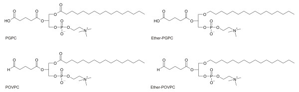
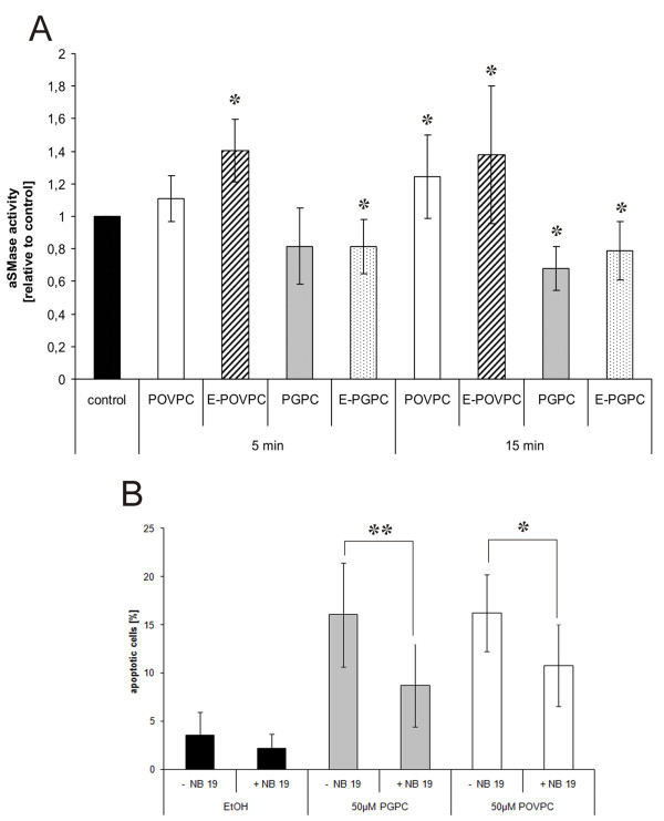
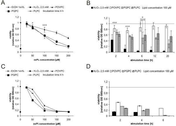
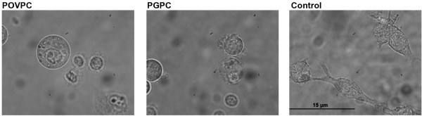
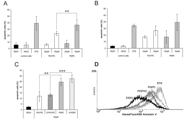
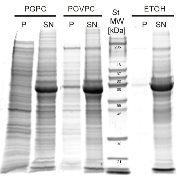
It was the aim of this study to determine the role of short-chain oxidized phospholipids as components of modified LDL in cultured macrophages. For this purpose we investigated the effects of the following oxidized phospholipids on cell viability and apoptosis: 1-palmitoyl-2-glutaroyl-sn-glycero-3-phosphocholine (PGPC), 1-palmitoyl-2-(5-oxovaleroyl)-sn-glycero-3-phosphocholine (POVPC) and oxidized alkylacyl phospholipids including 1-O-hexadecyl-2-glutaroyl-sn-glycero-3-phosphocholine (E-PGPC) and 1-O-hexadecyl-2-(5-oxovaleroyl)-sn-glycero-3-phosphocholine (E-POVPC). We found that these compounds induced apoptosis in RAW264.7 and bone marrow-derived macrophages. The sn-2 carboxyacyl lipid PGPC was more toxic than POVPC which carries a reactive aldehyde function in position sn-2 of glycerol. The alkylacyl phospholipids (E-PGPC and E-POVPC) and the respective diacyl analogs show similar activities. Apoptosis induced by POVPC and its alkylether derivative could be causally linked to the fast activation of an acid sphingomyelinase, generating the apoptotic second messenger ceramide. In contrast, PGPC and its ether analog only negligibly affected this enzyme pointing to an entirely different mechanism of lipid toxicity. The higher toxicity of PGPC is underscored by more efficient membrane blebbing from apoptotic cells. In addition, the protein pattern of PGPC-induced microparticles is different from the vesicles generated by POPVC.
In summary, our data reveal that oxidized phospholipids induce apoptosis in cultured macrophages. The mechanism of lipid toxicity, however, largely depends on the structural features of the oxidized sn-2 chain.
氧化型低密度脂蛋白(LDL)与巨噬细胞的相互作用是动脉粥样硬化发生发展的特征之一。修饰后的 LDL 颗粒在这些细胞中的生物学活性,归因于脂质氧化产物的含量和氧化磷脂对载脂蛋白的修饰。
本研究旨在确定短链氧化磷脂作为修饰型 LDL 中成分在培养的巨噬细胞中的作用。为此,我们研究了以下氧化磷脂对细胞活力和细胞凋亡的影响:1-棕榈酰基-2-戊二酰基-sn-甘油-3-磷酸胆碱(PGPC)、1-棕榈酰基-2-(5-氧戊酰基)-sn-甘油-3-磷酸胆碱(POVPC)以及氧化的烷基酰基磷脂,包括 1-O-十六烷基-2-戊二酰基-sn-甘油-3-磷酸胆碱(E-PGPC)和 1-O-十六烷基-2-(5-氧戊酰基)-sn-甘油-3-磷酸胆碱(E-POVPC)。我们发现,这些化合物诱导 RAW264.7 和骨髓来源的巨噬细胞发生细胞凋亡。甘油 sn-2 位羧酸酰基的 PGPC 比 POVPC 毒性更大,POVPC 在甘油 sn-2 位含有反应性醛基。烷基酰基磷脂(E-PGPC 和 E-POVPC)及其相应的二酰基类似物具有相似的活性。POVPC 及其醚衍生物诱导的细胞凋亡与酸性鞘磷脂酶的快速激活有关,从而产生凋亡的第二信使神经酰胺。相比之下,PGPC 及其醚类似物对该酶的影响可忽略不计,这表明脂质毒性的机制完全不同。PGPC 从凋亡细胞中更有效地形成质膜泡,这突显了其更高的毒性。此外,PGPC 诱导的微粒的蛋白质图谱与 POPVC 产生的囊泡不同。
综上所述,我们的数据表明氧化磷脂诱导培养的巨噬细胞发生细胞凋亡。然而,脂质毒性的机制在很大程度上取决于氧化 sn-2 链的结构特征。