Division of Radiation Cancer Biology, Korea Institute of Radiological and Medical Sciences, 215-4, Gongneung-Dong, Nowon-Gu, Seoul 139-706, Republic of Korea.
Radiat Oncol. 2012 Sep 11;7:153. doi: 10.1186/1748-717X-7-153.
Γ-Ionizing radiation (IR) therapy is one of major therapeutic tools in cancer treatment. Nevertheless, γ-IR therapy failed due to occurrence of metastasis, which constitutes a significant obstacle in cancer treatment. The main aim of this investigation was to construct animal model which present metastasis during radiotherapy in a mouse system in vivo and establishes the molecular mechanisms involved.
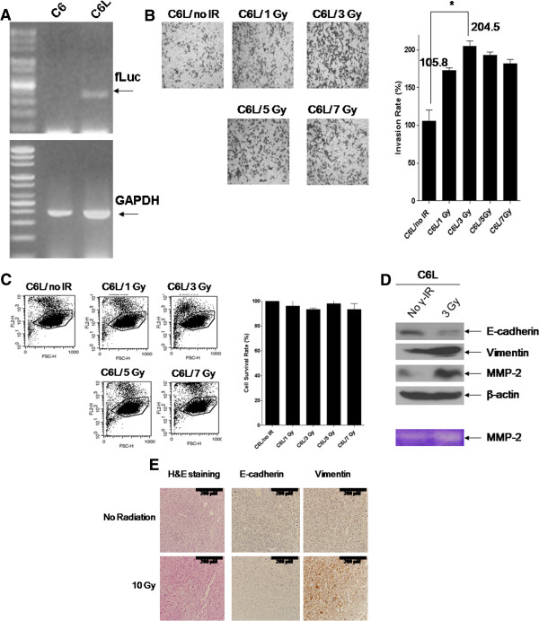
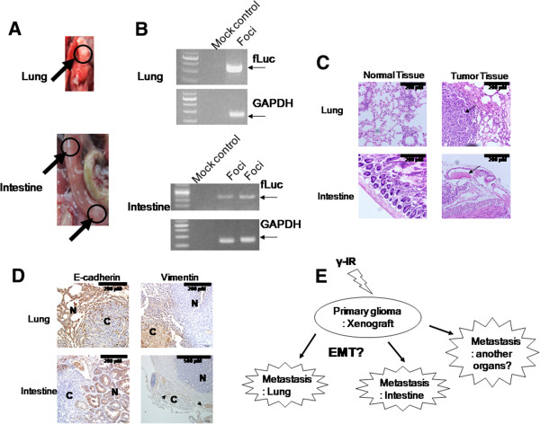
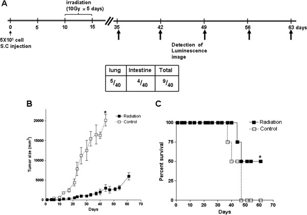
The C6L transfectant cell line expressing firefly luciferase (fLuc) was treated with γ-IR, followed by immunoblotting, zymography and invasion assay in vitro. We additionally employed the C6L transfectant cell line to construct xenografts in nude mice, which were irradiated with γ-IR. Irradiated xenograft-containing mice were analyzed via survival curves, measurement of tumor size, and bioluminescence imaging in vivo and ex vivo. Metastatic lesions in organs of mice were further assessed using RT-PCR, H & E staining and immunohistochemistry.
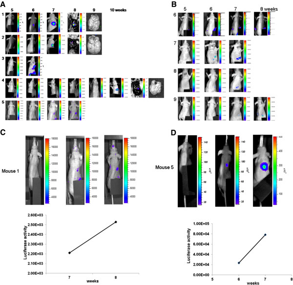
γ-IR treatment of C6L cells induced epithelial-mesenchymal transition (EMT) and increased cell invasion. In irradiated xenograft-containing mice, tumor sizes were decreased dramatically and survival rates extended. Almost all non-irradiated xenograft-containing control mice had died within 4 weeks. However, we also observed luminescence signals in about 22.5% of γ-IR-treated mice. Intestines or lungs of mice displaying luminescence signals contained several lesions, which expressed the fLuc gene and presented histological features of cancer tissues as well as expression of EMT markers.
These findings collectively indicate that occurrences of metastases during γ-IR treatment accompanied induction of EMT markers, including increased MMP activity. Establishment of a murine metastasis model during γ-IR treatment should aid in drug development against cancer metastasis and increase our understanding of the mechanisms underlying the metastatic process.
γ-离子化辐射(IR)治疗是癌症治疗的主要治疗工具之一。然而,由于转移的发生,γ-IR 治疗失败了,这是癌症治疗的一个重大障碍。本研究的主要目的是构建一种动物模型,在体内小鼠系统中表现出放射治疗期间的转移,并确定涉及的分子机制。
表达萤火虫荧光素酶(fLuc)的 C6L 转染细胞系用 γ-IR 处理,然后进行体外免疫印迹、酶谱分析和侵袭试验。我们还利用 C6L 转染细胞系在裸鼠中构建异种移植物,并用 γ-IR 照射。用 γ-IR 照射的异种移植物含有的小鼠通过生存曲线、肿瘤大小测量以及体内和体外生物发光成像进行分析。使用 RT-PCR、H & E 染色和免疫组织化学进一步评估小鼠器官中的转移病灶。
γ-IR 处理 C6L 细胞诱导上皮-间充质转化(EMT)并增加细胞侵袭。在照射的异种移植物含有的小鼠中,肿瘤大小显著减小,生存率延长。几乎所有未经照射的异种移植物含有的对照小鼠在 4 周内死亡。然而,我们也观察到约 22.5%的γ-IR 处理小鼠中出现了发光信号。显示发光信号的小鼠的肠道或肺部含有几个病灶,这些病灶表达 fLuc 基因,并表现出癌症组织的组织学特征以及 EMT 标志物的表达。
这些发现共同表明,γ-IR 治疗期间转移的发生伴随着 EMT 标志物的诱导,包括 MMP 活性的增加。在 γ-IR 治疗期间建立小鼠转移模型应有助于开发针对癌症转移的药物,并增加我们对转移过程中机制的理解。