Institut des Sciences Moléculaires d'Orsay, CNRS UMR 8214, Univ. Paris Sud, Orsay, France.
PLoS One. 2012;7(9):e44434. doi: 10.1371/journal.pone.0044434. Epub 2012 Sep 4.
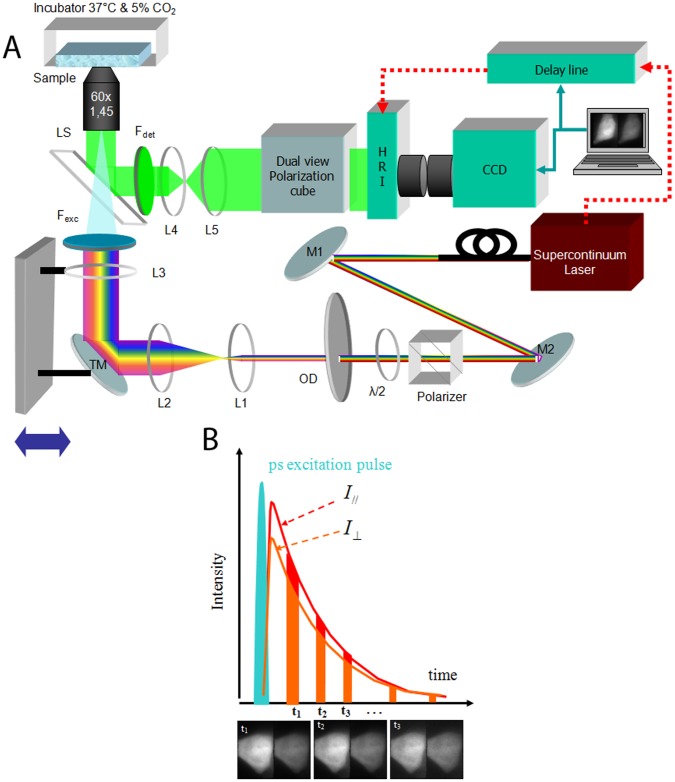
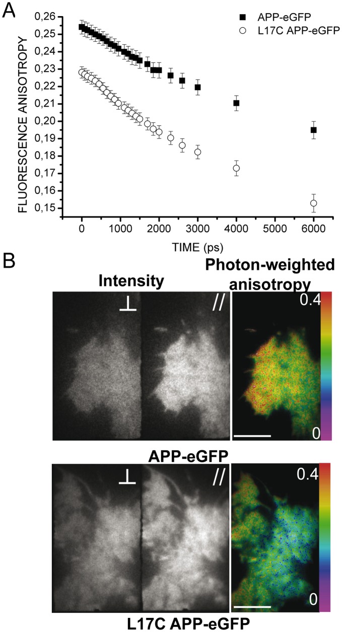
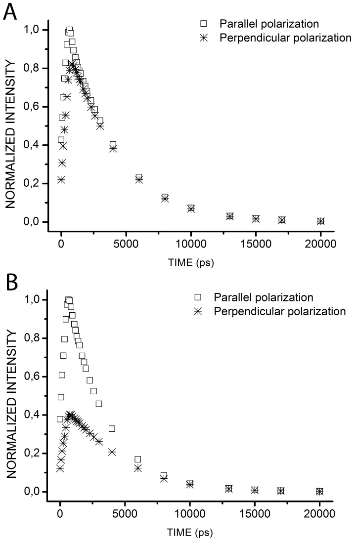
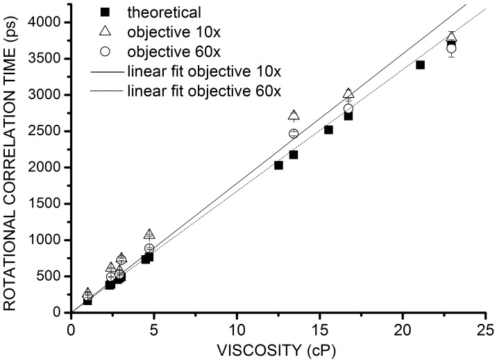
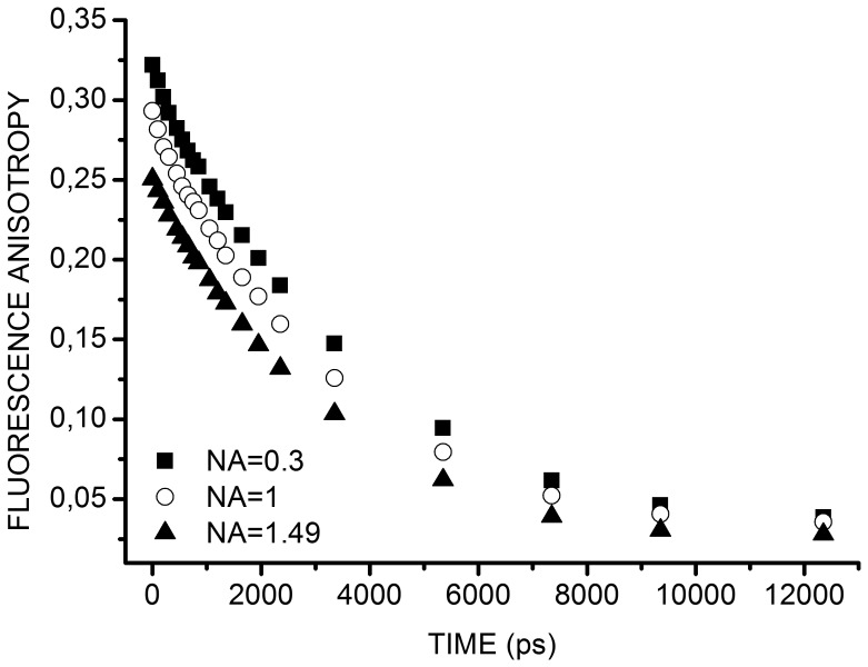
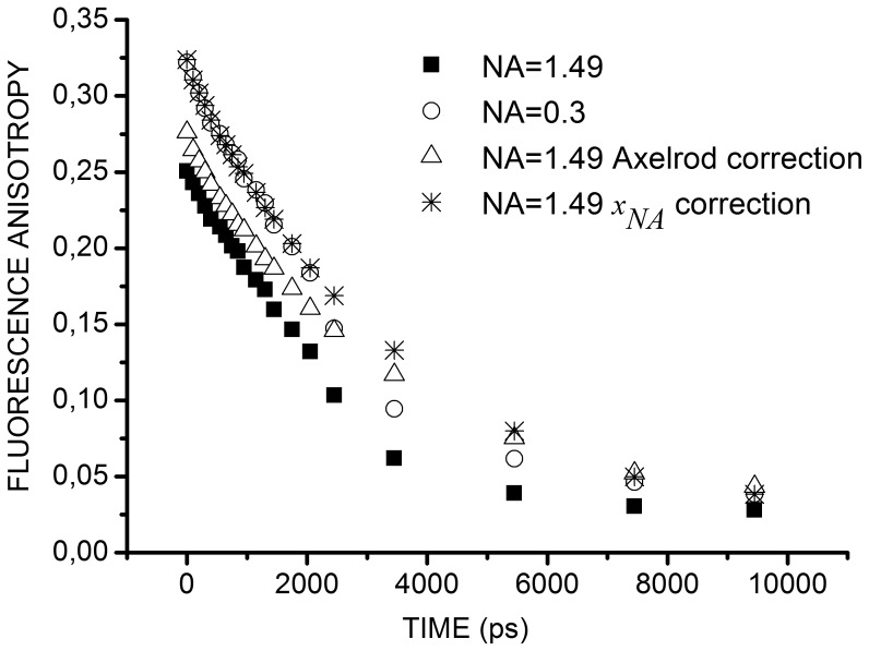
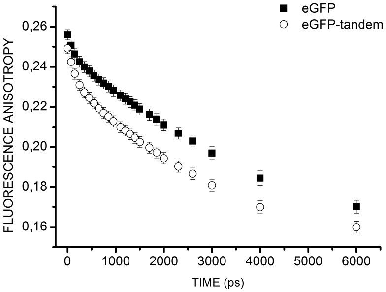
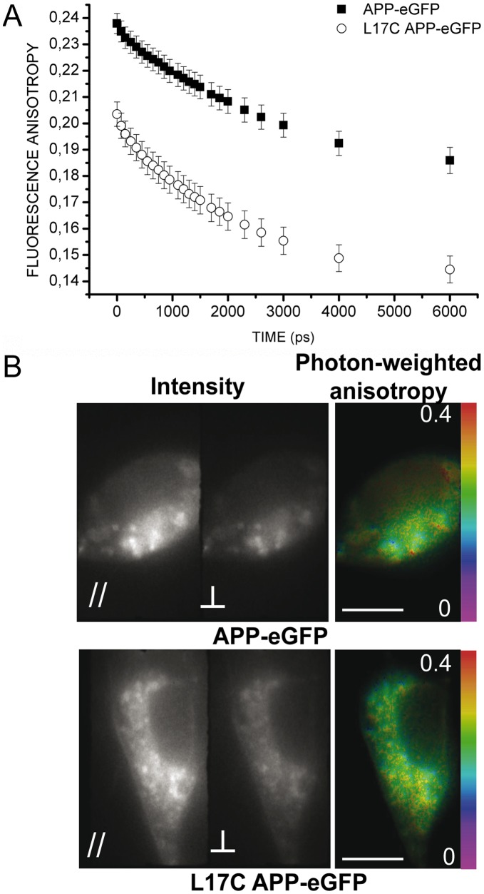
Classical FRET (Förster Resonance Energy Transfer) using two fluorescent labels (one for the donor and another one for the acceptor) is not efficient for studying the homodimerization of a protein as only half of the homodimers formed can be identified by this technique. We thus resorted to homoFRET detected by time-resolved Fluorescence Anisotropy IMaging (tr-FAIM). To specifically image the plasma membrane of living cells, an original combination of tr-FAIM and Total Internal Reflection Fluorescence Lifetime Imaging Microscope (TIRFLIM) was implemented. The correcting factor accounting for the depolarization due to the high numerical aperture (NA) objective, mandatory for TIRF microscopy, was quantified on fluorescein solutions and on HEK293 cells expressing enhanced Green Fluorescence Protein (eGFP). Homodimerization of Amyloid Precursor Protein (APP), a key mechanism in the etiology of Alzheimer's disease, was measured on this original set-up. We showed, both in epifluorescence and under TIRF excitation, different energy transfer rates associated with the homodimerization of wild type APP-eGFP or of a mutated APP-eGFP, which forms constitutive dimers. This original set-up thus offers promising prospects for future studies of protein homodimerization in living cells in control and pathological conditions.
经典的荧光共振能量转移(Förster Resonance Energy Transfer,FRET)技术使用两个荧光标记物(一个用于供体,另一个用于受体),对于研究蛋白质的同源二聚化并不有效,因为只有一半形成的同源二聚体可以通过这种技术来识别。因此,我们采用了通过时间分辨荧光各向异性成像(time-resolved Fluorescence Anisotropy IMaging,tr-FAIM)检测的同源 FRET。为了专门对活细胞的质膜进行成像,我们实现了 tr-FAIM 和全内反射荧光寿命成像显微镜(Total Internal Reflection Fluorescence Lifetime Imaging Microscope,TIRFLIM)的原始组合。由于 TIRF 显微镜需要高数值孔径(numerical aperture,NA)物镜,因此必须考虑由于高数值孔径引起的去极化,我们对荧光素溶液和表达增强型绿色荧光蛋白(enhanced Green Fluorescence Protein,eGFP)的 HEK293 细胞进行了定量。我们在这个原始设置上测量了阿尔茨海默病发病机制中的关键机制淀粉样前体蛋白(Amyloid Precursor Protein,APP)的同源二聚化。我们在明场和 TIRF 激发下都显示了与野生型 APP-eGFP 或组成型二聚体形成的突变型 APP-eGFP 同源二聚化相关的不同能量转移率。因此,这个原始设置为在生理和病理条件下研究活细胞中蛋白质同源二聚化提供了有前途的前景。