• 文献检索
  • 文档翻译
  • 深度研究
  • 学术资讯
  • Suppr Zotero 插件Zotero 插件
  • 邀请有礼
  • 套餐&价格
  • 历史记录
应用&插件
Suppr Zotero 插件Zotero 插件浏览器插件Mac 客户端Windows 客户端微信小程序
定价
高级版会员购买积分包购买API积分包
服务
文献检索文档翻译深度研究API 文档MCP 服务
关于我们
关于 Suppr公司介绍联系我们用户协议隐私条款
关注我们

Suppr 超能文献

核心技术专利:CN118964589B侵权必究
粤ICP备2023148730 号-1Suppr @ 2026

文献检索

告别复杂PubMed语法,用中文像聊天一样搜索,搜遍4000万医学文献。AI智能推荐,让科研检索更轻松。

立即免费搜索

文件翻译

保留排版,准确专业,支持PDF/Word/PPT等文件格式,支持 12+语言互译。

免费翻译文档

深度研究

AI帮你快速写综述,25分钟生成高质量综述,智能提取关键信息,辅助科研写作。

立即免费体验

使用 T2 弛豫定量法自动检测脑白质信号异常:在极早产儿足月 MRI 脑分割中的应用。

Automated detection of white matter signal abnormality using T2 relaxometry: application to brain segmentation on term MRI in very preterm infants.

机构信息

Center for Perinatal Research, The Research Institute at Nationwide Children's Hospital, Columbus, OH 43215, USA.

出版信息

Neuroimage. 2013 Jan 1;64:328-40. doi: 10.1016/j.neuroimage.2012.08.081. Epub 2012 Sep 6.

DOI:10.1016/j.neuroimage.2012.08.081
PMID:22974556
原文链接:https://pmc.ncbi.nlm.nih.gov/articles/PMC3544934/
Abstract

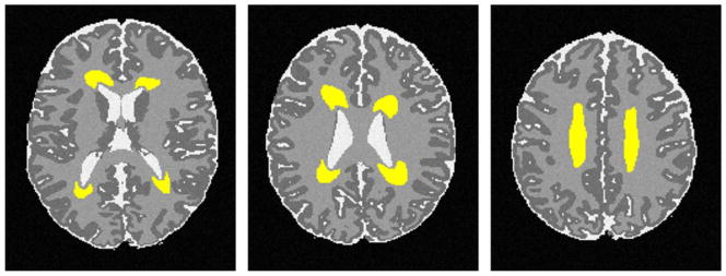
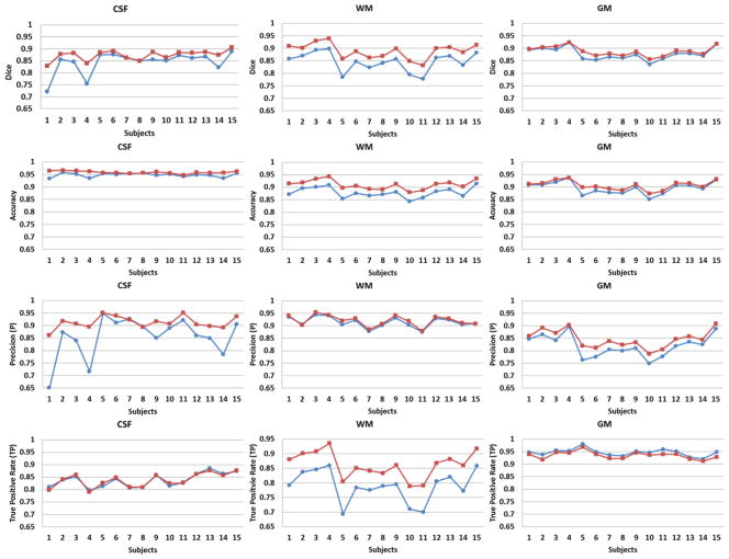
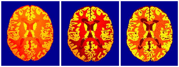
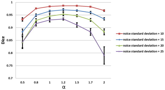
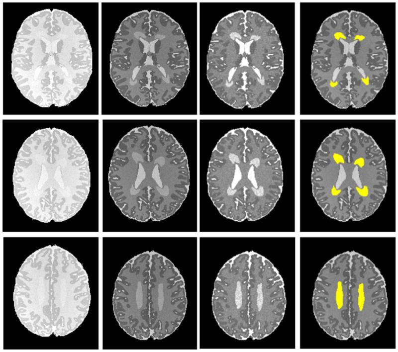
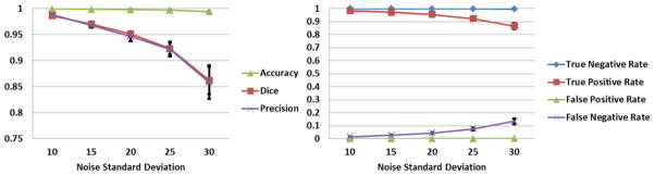
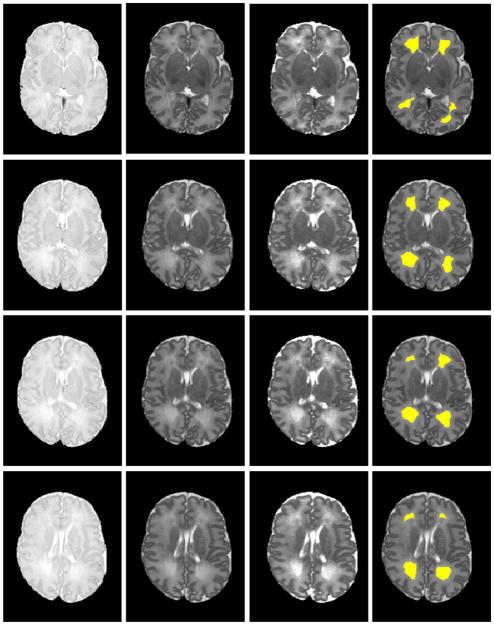
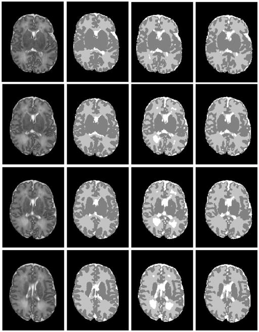
Hyperintense white matter signal abnormalities, also called diffuse excessive high signal intensity (DEHSI), are observed in up to 80% of very preterm infants on T2-weighted MRI scans at term-equivalent age. DEHSI may represent a developmental stage or diffuse microstructural white matter abnormalities. Automated quantitative assessment of DEHSI severity may help resolve this debate and improve neonatal brain tissue segmentation. For T2-weighted sequence without fluid attenuation, the signal intensity distribution of DEHSI greatly overlaps with that of cerebrospinal fluid (CSF) making its detection difficult. Furthermore, signal intensities of T2-weighted images are susceptible to magnetic field inhomogeneity. Increased signal intensities caused by field inhomogeneity may be confused with DEHSI. To overcome these challenges, we propose an algorithm to detect DEHSI using T2 relaxometry, whose reflection of the rapid changes in free water content provides improved distinction between CSF and DEHSI over that of conventional T2-weighted imaging. Moreover, the parametric transverse relaxation time T2 is invulnerable to magnetic field inhomogeneity. We conducted computer simulations to select an optimal detection parameter and to validate the proposed method. We also demonstrated that brain tissue segmentation is further enhanced by incorporating DEHSI detection for both simulated preterm infant brain images and in vivo in very preterm infants imaged at term-equivalent age.

摘要

在足月时的磁共振成像扫描中,高达 80%的极早产儿可观察到高信号白质信号异常,也称为弥漫性过度高信号强度(DEHSI)。DEHSI 可能代表一种发育阶段或弥漫性的微观结构白质异常。DEHSI 严重程度的自动定量评估可能有助于解决这一争议并改善新生儿脑组织分割。对于没有液体衰减的 T2 加权序列,DEHSI 的信号强度分布与脑脊液(CSF)的信号强度分布有很大的重叠,这使得其检测变得困难。此外,T2 加权图像的信号强度容易受到磁场不均匀性的影响。由磁场不均匀性引起的信号强度增加可能与 DEHSI 混淆。为了克服这些挑战,我们提出了一种使用 T2 弛豫度来检测 DEHSI 的算法,其对游离水含量快速变化的反映提供了比传统 T2 加权成像更好的 CSF 和 DEHSI 之间的区分。此外,参数横向弛豫时间 T2 不受磁场不均匀性的影响。我们进行了计算机模拟,以选择最佳的检测参数并验证所提出的方法。我们还证明,通过将 DEHSI 检测纳入模拟早产儿脑图像和足月时成像的极早产儿的体内,脑组织分割得到了进一步增强。

https://cdn.ncbi.nlm.nih.gov/pmc/blobs/3ab1/3544934/3261ed0218ac/nihms405660f8a.jpg
https://cdn.ncbi.nlm.nih.gov/pmc/blobs/3ab1/3544934/ac8428a3c69d/nihms405660f1.jpg
https://cdn.ncbi.nlm.nih.gov/pmc/blobs/3ab1/3544934/c7696be84a34/nihms405660f2.jpg
https://cdn.ncbi.nlm.nih.gov/pmc/blobs/3ab1/3544934/a77e0a98f6f3/nihms405660f3.jpg
https://cdn.ncbi.nlm.nih.gov/pmc/blobs/3ab1/3544934/b7a55c17c7ab/nihms405660f4.jpg
https://cdn.ncbi.nlm.nih.gov/pmc/blobs/3ab1/3544934/4fcd149286e5/nihms405660f5.jpg
https://cdn.ncbi.nlm.nih.gov/pmc/blobs/3ab1/3544934/0cadaf3ac8e1/nihms405660f6a.jpg
https://cdn.ncbi.nlm.nih.gov/pmc/blobs/3ab1/3544934/21c90946fa98/nihms405660f7a.jpg
https://cdn.ncbi.nlm.nih.gov/pmc/blobs/3ab1/3544934/3261ed0218ac/nihms405660f8a.jpg

相似文献

1
Automated detection of white matter signal abnormality using T2 relaxometry: application to brain segmentation on term MRI in very preterm infants.使用 T2 弛豫定量法自动检测脑白质信号异常:在极早产儿足月 MRI 脑分割中的应用。
Neuroimage. 2013 Jan 1;64:328-40. doi: 10.1016/j.neuroimage.2012.08.081. Epub 2012 Sep 6.
2
T2 at MR imaging is an objective quantitative measure of cerebral white matter signal intensity abnormality in preterm infants at term-equivalent age.磁共振成像中的T2是足月等效年龄早产儿脑白质信号强度异常的客观定量指标。
Radiology. 2009 Jul;252(1):209-17. doi: 10.1148/radiol.2522080589.
3
Assessment of brain maturation in the preterm infants using diffusion tensor imaging (DTI) and enhanced T2 star weighted angiography (ESWAN).应用弥散张量成像(DTI)和增强 T2*星加权血管造影(ESWAN)评估早产儿脑成熟度。
Eur J Radiol. 2013 Sep;82(9):e476-83. doi: 10.1016/j.ejrad.2013.04.003. Epub 2013 Apr 29.
4
Diffusion-weighted imaging of the brain in preterm infants with focal and diffuse white matter abnormality.早产儿局灶性和弥漫性白质异常的脑扩散加权成像
Pediatrics. 2003 Jul;112(1 Pt 1):1-7. doi: 10.1542/peds.112.1.1.
5
Neuro-developmental outcome at 18 months in premature infants with diffuse excessive high signal intensity on MR imaging of the brain.脑弥散性过度高信号早产儿 18 个月时的神经发育结局的磁共振成像研究。
Pediatr Radiol. 2011 Oct;41(10):1284-92. doi: 10.1007/s00247-011-2155-7. Epub 2011 Jun 17.
6
White matter changes in extremely preterm infants, a population-based diffusion tensor imaging study.极早产儿脑白质改变:基于人群的弥散张量成像研究。
Acta Paediatr. 2010 Jun;99(6):842-9. doi: 10.1111/j.1651-2227.2009.01634.x. Epub 2010 Feb 1.
7
Application of variable threshold intensity to segmentation for white matter hyperintensities in fluid attenuated inversion recovery magnetic resonance images.可变阈值强度在液体衰减反转恢复磁共振图像中对白质高信号进行分割的应用。
Neuroradiology. 2014 Apr;56(4):265-81. doi: 10.1007/s00234-014-1322-6. Epub 2014 Feb 4.
8
The challenge of cerebral magnetic resonance imaging in neonates: A new method using mathematical morphology for the segmentation of structures including diffuse excessive high signal intensities.新生儿脑磁共振成像的挑战:一种新方法,使用数学形态学对包括弥漫性高信号强度在内的结构进行分割。
Med Image Anal. 2018 Aug;48:75-94. doi: 10.1016/j.media.2018.05.003. Epub 2018 May 17.
9
Diffuse excessive high signal intensity in low-risk preterm infants at term-equivalent age does not predict outcome at 1 year: a prospective study.足月相当年龄的低风险早产儿弥漫性过高信号强度不能预测1岁时的预后:一项前瞻性研究。
Neuroradiology. 2014 Aug;56(8):669-78. doi: 10.1007/s00234-014-1373-8. Epub 2014 May 14.
10
Diffusion MRI of the neonate brain: acquisition, processing and analysis techniques.新生儿脑弥散 MRI:采集、处理和分析技术。
Pediatr Radiol. 2012 Oct;42(10):1169-82. doi: 10.1007/s00247-012-2427-x. Epub 2012 Aug 18.

引用本文的文献

1
Duration of mechanical ventilation is more critical for brain growth than postnatal hydrocortisone in extremely preterm infants.机械通气时间对极早产儿脑生长的影响比出生后氢化可的松更为关键。
Eur J Pediatr. 2021 Nov;180(11):3307-3315. doi: 10.1007/s00431-021-04113-z. Epub 2021 May 16.
2
Perinatal Risk and Protective Factors in the Development of Diffuse White Matter Abnormality on Term-Equivalent Age Magnetic Resonance Imaging in Infants Born Very Preterm.极早产儿足月龄时磁共振弥散加权成像显示脑白质病变的围生期风险和保护因素。
J Pediatr. 2021 Jun;233:58-65.e3. doi: 10.1016/j.jpeds.2020.11.058. Epub 2020 Nov 28.
3

本文引用的文献

1
Qualitative brain MRI at term and cognitive outcomes at 9 years after very preterm birth.足月时的定性脑 MRI 与极早早产儿出生 9 年后的认知结局。
Pediatrics. 2012 May;129(5):e1138-47. doi: 10.1542/peds.2011-1735. Epub 2012 Apr 23.
2
Automated quantification of white matter disease extent at 3 T: comparison with volumetric readings.3T 下脑白质病变程度的自动量化:与容积读数的比较。
J Magn Reson Imaging. 2012 Aug;36(2):305-11. doi: 10.1002/jmri.23659. Epub 2012 Apr 19.
3
Neurodevelopmental outcomes in preterm infants: comparison of infants with and without diffuse excessive high signal intensity on MR images at near-term-equivalent age.
Objectively Diagnosed Diffuse White Matter Abnormality at Term Is an Independent Predictor of Cognitive and Language Outcomes in Infants Born Very Preterm.
足月时客观诊断的弥漫性脑白质异常是极早产儿认知和语言结局的独立预测因素。
J Pediatr. 2020 May;220:56-63. doi: 10.1016/j.jpeds.2020.01.034. Epub 2020 Mar 5.
4
Antecedents of Objectively Diagnosed Diffuse White Matter Abnormality in Very Preterm Infants.早产儿客观诊断的弥散性脑白质异常的相关因素。
Pediatr Neurol. 2020 May;106:56-62. doi: 10.1016/j.pediatrneurol.2020.01.011. Epub 2020 Feb 4.
5
Objective and Automated Detection of Diffuse White Matter Abnormality in Preterm Infants Using Deep Convolutional Neural Networks.使用深度卷积神经网络对早产儿弥漫性白质异常进行客观自动检测。
Front Neurosci. 2019 Jun 18;13:610. doi: 10.3389/fnins.2019.00610. eCollection 2019.
6
Thirteen-Year Outcomes in Very Preterm Children Associated with Diffuse Excessive High Signal Intensity on Neonatal Magnetic Resonance Imaging.新生儿磁共振弥散过度高信号与极早产儿 13 年结局的相关性。
J Pediatr. 2019 Mar;206:66-71.e1. doi: 10.1016/j.jpeds.2018.10.016. Epub 2018 Nov 7.
7
Optimization of magnetization-prepared rapid gradient echo (MP-RAGE) sequence for neonatal brain MRI.用于新生儿脑部磁共振成像的磁化准备快速梯度回波(MP-RAGE)序列的优化
Pediatr Radiol. 2018 Aug;48(8):1139-1151. doi: 10.1007/s00247-018-4140-x. Epub 2018 May 2.
8
T2 Relaxometry MRI Predicts Cerebral Palsy in Preterm Infants.T2 弛豫率 MRI 预测早产儿脑瘫。
AJNR Am J Neuroradiol. 2018 Mar;39(3):563-568. doi: 10.3174/ajnr.A5501. Epub 2018 Jan 18.
9
Differentiating T2 hyperintensity in neonatal white matter by two-compartment model of diffusional kurtosis imaging.通过扩散峰度成像的双室模型鉴别新生儿白质中的T2高信号。
Sci Rep. 2016 Apr 14;6:24473. doi: 10.1038/srep24473.
10
Longitudinal development in the preterm thalamus and posterior white matter: MRI correlations between diffusion weighted imaging and T2 relaxometry.早产儿丘脑和后白质的纵向发育:扩散加权成像与T2弛豫测量法之间的MRI相关性
Hum Brain Mapp. 2016 Jul;37(7):2479-92. doi: 10.1002/hbm.23188. Epub 2016 Mar 21.
早产儿的神经发育结局:在近足月龄时,比较磁共振成像上有无弥漫性过度高信号强度的早产儿。
Radiology. 2012 May;263(2):518-26. doi: 10.1148/radiol.12111615. Epub 2012 Mar 8.
4
Multi-stage segmentation of white matter hyperintensity, cortical and lacunar infarcts.脑白质高信号、皮质及腔隙性梗死的多阶段分割。
Neuroimage. 2012 May 1;60(4):2379-88. doi: 10.1016/j.neuroimage.2012.02.034. Epub 2012 Feb 22.
5
Brain tissue volumes in preterm infants: prematurity, perinatal risk factors and neurodevelopmental outcome: a systematic review.早产儿的脑组织体积:早产、围产期危险因素及神经发育结局:一项系统综述
J Matern Fetal Neonatal Med. 2012 Apr;25 Suppl 1:89-100. doi: 10.3109/14767058.2012.664343.
6
Automated measurement of local white matter lesion volume.自动测量局部脑白质病变体积。
Neuroimage. 2012 Feb 15;59(4):3901-8. doi: 10.1016/j.neuroimage.2011.11.021. Epub 2011 Nov 11.
7
Clinical implications of MR imaging findings in the white matter in very preterm infants: a 2-year follow-up study.早产儿脑白质磁共振成像表现的临床意义:一项 2 年随访研究。
Radiology. 2011 Dec;261(3):899-906. doi: 10.1148/radiol.11110797. Epub 2011 Oct 26.
8
High signal intensity on T2-weighted MR imaging at term-equivalent age in preterm infants does not predict 2-year neurodevelopmental outcomes.足月龄等龄早产儿 T2 加权磁共振成像高信号强度不能预测 2 年神经发育结局。
AJNR Am J Neuroradiol. 2011 Dec;32(11):2005-10. doi: 10.3174/ajnr.A2703. Epub 2011 Sep 29.
9
Automatic segmentation of neonatal images using convex optimization and coupled level sets.基于凸优化和耦合水平集的新生儿图像自动分割。
Neuroimage. 2011 Oct 1;58(3):805-17. doi: 10.1016/j.neuroimage.2011.06.064. Epub 2011 Jul 5.
10
Human brain iron mapping using atlas-based T2 relaxometry.基于图谱的 T2 弛豫率法人脑铁定量成像
Magn Reson Med. 2012 Mar;67(3):731-9. doi: 10.1002/mrm.23054. Epub 2011 Jun 23.