Department of Clinical and Experimental Epilepsy, UCL Institute of Neurology, Queen Square, London WC1N 3BG, UK.
Brain. 2012 Oct;135(Pt 10):3101-14. doi: 10.1093/brain/aws232. Epub 2012 Sep 13.
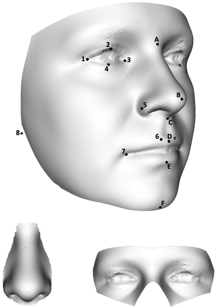
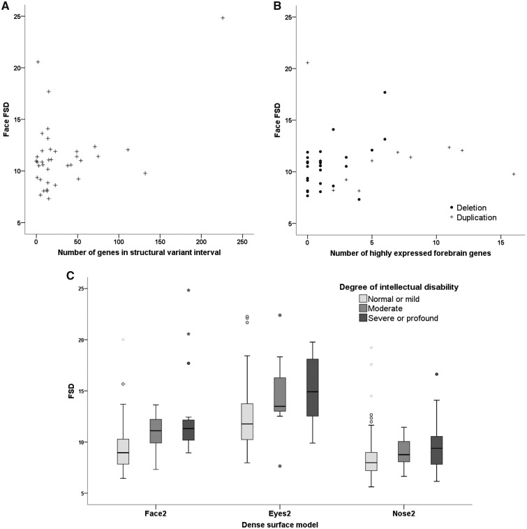
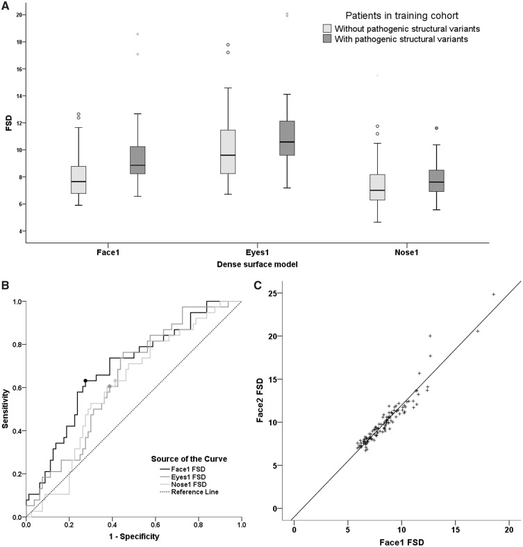
Many pathogenic structural variants of the human genome are known to cause facial dysmorphism. During the past decade, pathogenic structural variants have also been found to be an important class of genetic risk factor for epilepsy. In other fields, face shape has been assessed objectively using 3D stereophotogrammetry and dense surface models. We hypothesized that computer-based analysis of 3D face images would detect subtle facial abnormality in people with epilepsy who carry pathogenic structural variants as determined by chromosome microarray. In 118 children and adults attending three European epilepsy clinics, we used an objective measure called Face Shape Difference to show that those with pathogenic structural variants have a significantly more atypical face shape than those without such variants. This is true when analysing the whole face, or the periorbital region or the perinasal region alone. We then tested the predictive accuracy of our measure in a second group of 63 patients. Using a minimum threshold to detect face shape abnormalities with pathogenic structural variants, we found high sensitivity (4/5, 80% for whole face; 3/5, 60% for periorbital and perinasal regions) and specificity (45/58, 78% for whole face and perinasal regions; 40/58, 69% for periorbital region). We show that the results do not seem to be affected by facial injury, facial expression, intellectual disability, drug history or demographic differences. Finally, we use bioinformatics tools to explore relationships between facial shape and gene expression within the developing forebrain. Stereophotogrammetry and dense surface models are powerful, objective, non-contact methods of detecting relevant face shape abnormalities. We demonstrate that they are useful in identifying atypical face shape in adults or children with structural variants, and they may give insights into the molecular genetics of facial development.
已知许多人类基因组的致病性结构变体可导致面部畸形。在过去的十年中,致病性结构变体也被发现是癫痫的一个重要遗传风险因素类别。在其他领域,已经使用 3D 立体摄影测量术和密集表面模型来客观评估脸型。我们假设,通过计算机对面部 3D 图像进行分析,可以检测出携带染色体微阵列确定的致病性结构变体的癫痫患者的微妙面部异常。在参加三个欧洲癫痫诊所的 118 名儿童和成年人中,我们使用了一种称为“面部形状差异”的客观测量方法来表明,患有致病性结构变体的人具有明显更不典型的面部形状,而没有这些变体的人则没有。这是在分析整个面部、眶周区域或鼻周区域时的情况。然后,我们在第二组 63 名患者中测试了我们的测量方法的预测准确性。使用最小阈值来检测致病性结构变体引起的面部形状异常,我们发现了高灵敏度(整个面部为 4/5,80%;眶周和鼻周区域为 3/5,60%)和特异性(整个面部和鼻周区域为 45/58,78%;眶周区域为 40/58,69%)。我们表明,结果似乎不受面部损伤、面部表情、智力障碍、药物史或人口统计学差异的影响。最后,我们使用生物信息学工具探索面部形状与发育中前脑基因表达之间的关系。立体摄影测量术和密集表面模型是检测相关面部形状异常的强大、客观、非接触方法。我们证明,它们可用于识别患有结构变体的成年人或儿童的非典型面部形状,并且它们可能为面部发育的分子遗传学提供见解。