Surgical Neurology Branch, National Institute of Neurological Disorders and Stroke, National Institutes of Health, Bethesda, Maryland 20892-1414, USA.
J Neurosurg. 2012 Nov;117(5):942-6. doi: 10.3171/2012.8.JNS111476. Epub 2012 Sep 14.
Chiari malformation Type I (CM-I) is characterized by hindbrain deformity. We investigated the effects of craniocervical decompression surgery on the anatomical features of hindbrain deformity with a prospective MRI study of patients with CM-I.
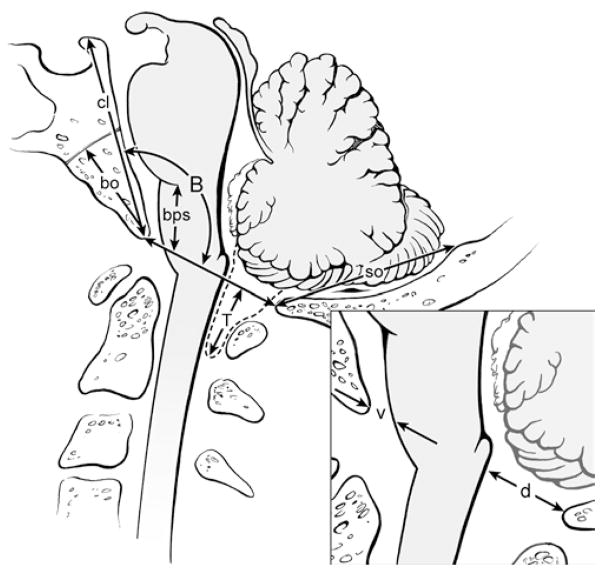
A prospective longitudinal study was conducted in 48 patients with CM-I (39 with syringomyelia) treated with craniocervical decompression. Clinical examinations and cervical MRI were performed before surgery and 1 week, 3-6 months, and annually after surgery. Hindbrain deformity was defined by tonsillar ectopia, pointed cerebellar tonsils, and/or cervicomedullary protuberance. The length of the clivus, basiocciput (sphenooccipital synchondrosis to basion), supraocciput (internal occipital protuberance to opisthion), and anteroposterior (AP) width of CSF pathways at the foramen magnum were measured and compared with those from 18 healthy volunteers (control group).
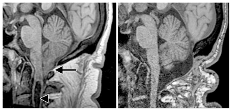
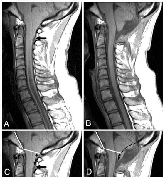
Before surgery, the patients' posterior fossa bones were short and their CSF pathways were narrow. All patients had tonsillar ectopia (mean [± SD] 12.3 ± 5.1 mm; normal 0.3 ± 1.0). The majority of patients had pointed tonsils and more than two-thirds exhibited a cervicomedullary protuberance. Clivus and basiocciput lengths were significantly shorter than the values obtained in the control group. However, the supraocciput length did not differ significantly from control measurements. The mean bulbopontine sulcus distance superior to the basion was 9.5 ± 2.6 mm (vs 13.6 ± 2.8 mm in controls; p < 0.0001). The AP widths of the CSF pathways at the level of the foramen magnum were significantly narrowed. After surgery, CSF pathways significantly expanded both ventrally and dorsally. By 3-6 months after surgery, pointed tonsils became round, cervicomedullary protuberance disappeared, and tonsillar ectopia diminished by 51% (to 6.0 ± 3.3 mm; p < 0.0001).
The cerebellar tonsils and brainstem assumed a normal appearance within 6 months after craniocervical decompression. These findings support the concept that the CM-I is not a congenital malformation of the neural elements but rather an acquired malformation that arises from pulsatile impaction of the cerebellar tonsils into the foramen magnum. Clinical trial registration no.: NCT00001327.
Chiari 畸形 I 型(CM-I)的特征是后脑畸形。我们通过前瞻性 MRI 研究对伴有 CM-I 的患者进行颅颈减压手术对后脑畸形解剖特征的影响。
对 48 例 CM-I(39 例伴有脊髓空洞症)患者进行前瞻性纵向研究,这些患者接受颅颈减压治疗。手术前及手术后 1 周、3-6 个月及每年进行临床检查和颈椎 MRI。后脑畸形定义为扁桃体下移、小脑扁桃体变尖和/或颈髓突出。测量并比较颅底(蝶枕结合处至基线)、颅顶(内枕骨结节至枕骨大孔)和矢状位(大孔内 CSF 通路的前后径)的斜坡长度、基底部(蝶枕结合处至基线)、颅顶(内枕骨结节至枕骨大孔)和矢状位(大孔内 CSF 通路的前后径),并与 18 名健康志愿者(对照组)进行比较。
手术前,患者的后颅窝骨骼短小,CSF 通路狭窄。所有患者均有扁桃体下移(平均[±SD]12.3±5.1mm;正常范围 0.3±1.0mm)。大多数患者的小脑扁桃体变尖,超过三分之二的患者出现颈髓突出。斜坡和颅底长度明显短于对照组。然而,颅顶长度与对照组无显著差异。基底结节上方的脑桥延髓沟距离为 9.5±2.6mm(对照组为 13.6±2.8mm;p<0.0001)。大孔水平 CSF 通路的前后径明显变窄。手术后,CSF 通路在腹侧和背侧均显著扩张。术后 3-6 个月,小脑扁桃体变圆,颈髓突出消失,扁桃体下移减少 51%(至 6.0±3.3mm;p<0.0001)。
小脑扁桃体和脑干在颅颈减压后 6 个月内恢复正常外观。这些发现支持 Chiari 畸形不是神经元素的先天性畸形,而是一种获得性畸形,源于小脑扁桃体搏动性冲击大孔。临床试验注册号:NCT00001327。