IPHT - Institute for Photonic Technology, Albert-Einstein-Strasse 9, D-07745 Jena, Germany.
Beilstein J Nanotechnol. 2012;3:404-14. doi: 10.3762/bjnano.3.47. Epub 2012 May 18.
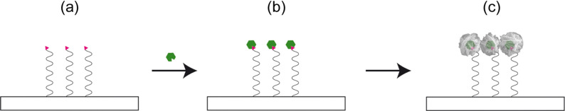
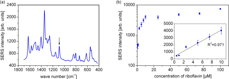
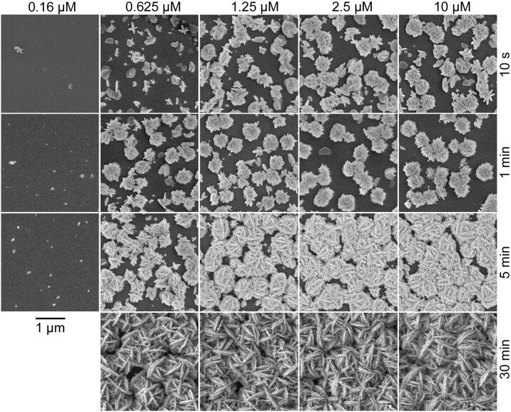
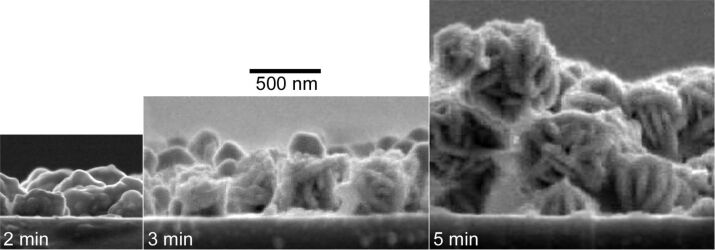
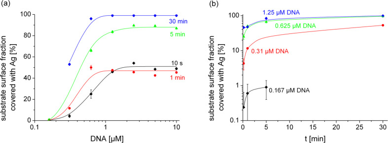
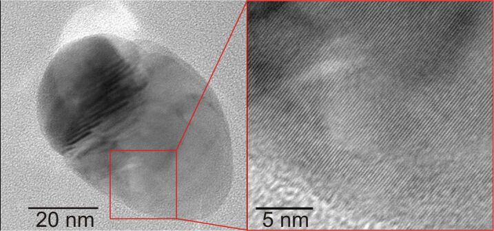
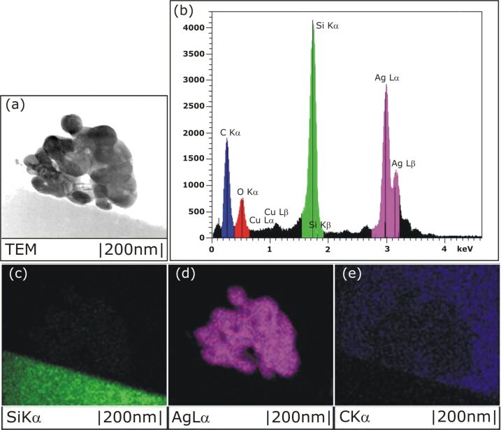
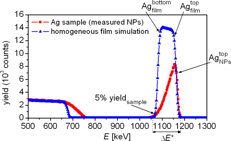
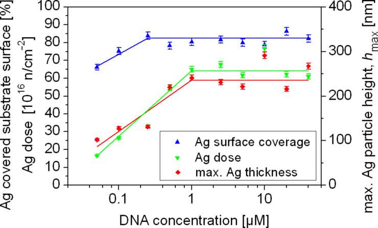
Silver nanoparticles were synthesized by an enzyme-induced growth process on solid substrates. In order to customize the enzymatically grown nanoparticles (EGNP) for analytical applications in biomolecular research, a detailed study was carried out concerning the time evolution of the formation of the silver nanoparticles, their morphology, and their chemical composition. Therefore, silver-nanoparticle films of different densities were investigated by using scanning as well as transmission electron microscopy to examine their structure. Cross sections of silver nanoparticles, prepared for analysis by transmission electron microscopy were additionally studied by energy-dispersive X-ray spectroscopy in order to probe their chemical composition. The surface coverage of substrates with silver nanoparticles and the maximum particle height were determined by Rutherford backscattering spectroscopy. Variations in the silver-nanoparticle films depending on the conditions during synthesis were observed. After an initial growth state the silver nanoparticles exhibit the so-called desert-rose or nanoflower-like structure. This complex nanoparticle structure is in clear contrast to the auto-catalytically grown spherical particles, which maintain their overall geometrical appearance while increasing their diameter. It is shown, that the desert-rose-like silver nanoparticles consist of single-crystalline plates of pure silver. The surface-enhanced Raman spectroscopic (SERS) activity of the EGNP structures is promising due to the exceptionally rough surface structure of the silver nanoparticles. SERS measurements of the vitamin riboflavin incubated on the silver nanoparticles are shown as an exemplary application for quantitative analysis.
银纳米粒子通过在固体基底上的酶诱导生长过程合成。为了将酶促生长的纳米粒子(EGNP)定制用于生物分子研究中的分析应用,对银纳米粒子的形成、形态和化学组成的时间演变进行了详细研究。因此,使用扫描和透射电子显微镜研究了不同密度的银纳米粒子薄膜,以检查它们的结构。此外,通过能量色散 X 射线光谱法对用于透射电子显微镜分析的银纳米粒子的横截面进行了研究,以探测其化学组成。通过卢瑟福背散射光谱法确定了具有银纳米粒子的基底的表面覆盖率和最大颗粒高度。观察到银纳米粒子薄膜随合成条件变化而变化。在初始生长状态之后,银纳米粒子表现出所谓的沙漠玫瑰或纳米花状结构。这种复杂的纳米粒子结构与自催化生长的球形粒子形成鲜明对比,后者在增加直径的同时保持其整体几何形状。结果表明,沙漠玫瑰状银纳米粒子由纯银的单晶片组成。由于银纳米粒子的异常粗糙表面结构,其表面增强拉曼光谱(SERS)活性很有前途。作为定量分析的示例应用,展示了在银纳米粒子上孵育的维生素核黄素的 SERS 测量结果。