Department of Pathology, Immunology and Laboratory Medicine, University of Florida College of Medicine, Gainesville, Florida 32610, USA.
J Urol. 2013 Mar;189(3):803-11. doi: 10.1016/j.juro.2012.05.078. Epub 2012 Sep 25.
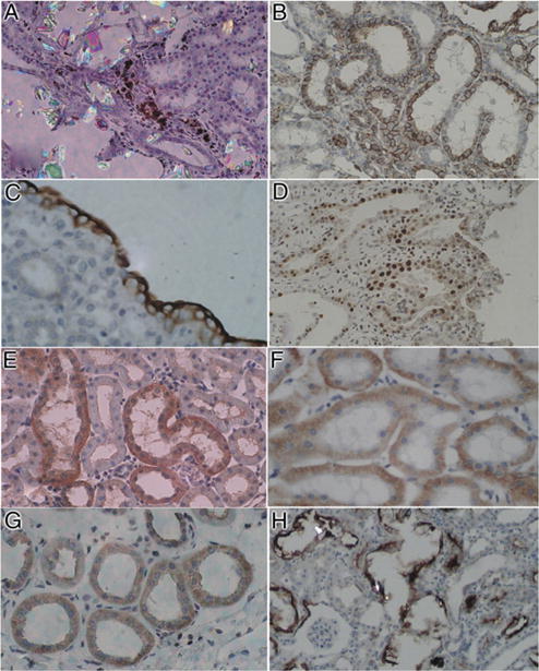
Idiopathic calcium oxalate kidney stones form while attached to Randall plaques, the subepithelial deposits on renal papillary surfaces. Plaque formation and growth mechanisms are poorly understood. Plaque formation elsewhere in the body is triggered by reactive oxygen species and oxidative stress. This review explores possible reactive oxygen species involvement in plaque formation and calcium oxalate nephrolithiasis.
A search of various databases for the last 8 years identified literature on reactive oxygen species involvement in calcium oxalate nephrolithiasis. The literature was reviewed and results are discussed.
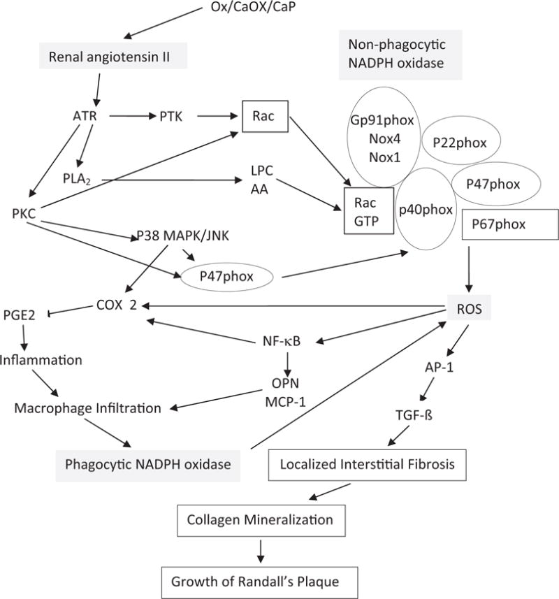
Under normal conditions reactive oxygen species production is controlled, increasing as needed and regulating crystallization modulator production. Reactive oxygen species overproduction or decreased antioxidants lead to oxidative stress, inflammation and injury, and are involved in stone comorbidity. All major chronic inflammation markers are detectable in stone patient urine. Patients also have increased urinary excretion of the IαI and the thrombin protein families. Results of a recent study of 17,695 participants in NHANES III (National Health and Nutrition Examination Survey) showed significantly lower antioxidants, carotene and β-cryptoxanthin in those with a kidney stone history. Animal model and tissue culture studies revealed that high oxalate, calcium oxalate and calcium phosphate crystals provoked renal cell reactive oxygen species mediated inflammatory responses. Calcium oxalate crystals induce renin up-regulation and angiotensin II generation. Nonphagocytic NADPH oxidase leads to reactive oxygen species production mediated by protein kinase C. The P-38 MAPK/JNK transduction pathway is turned on. Transcriptional and growth factors, and generated secondary mediators become involved. Chemoattractant and osteopontin production is increased and macrophages infiltrate the renal interstitium around the crystal. Phagocytic NADPH oxidase is probably activated, producing additional reactive oxygen species. Localized inflammation, extracellular matrix and fibrosis develop. Crystallization modulators have a significant role in inflammation and tissue repair.
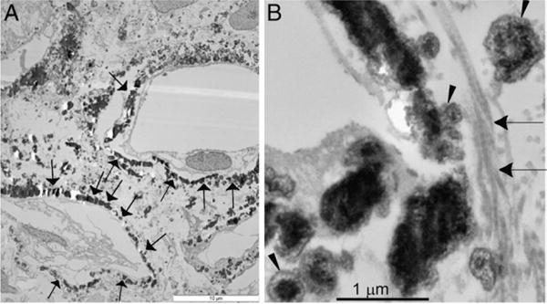
Based on available data, Randall plaque formation is similar to extracellular matrix mineralization at many body sites. Renal interstitial collagen becomes mineralized, assisting plaque growth through the interstitium until the mineralizing front reaches papillary surface epithelium. Plaque exposure to pelvic urine may also be a result of reactive oxygen species triggered epithelial sloughing.
特发性草酸钙肾结石形成于肾乳头表面的上皮下沉积物 Randall 斑上。斑块形成和生长的机制尚不清楚。身体其他部位的斑块形成是由活性氧和氧化应激引发的。本综述探讨了活性氧在斑块形成和草酸钙肾结石中的可能作用。
在过去 8 年中,通过对各种数据库的搜索,确定了有关活性氧参与草酸钙肾结石的文献。对文献进行了回顾和讨论。
在正常情况下,活性氧的产生受到控制,根据需要增加,并调节结晶调节剂的产生。活性氧的过度产生或抗氧化剂的减少会导致氧化应激、炎症和损伤,并与结石的合并症有关。所有主要的慢性炎症标志物都可在结石患者的尿液中检测到。患者的尿液中还存在 IαI 和凝血酶蛋白家族的排泄增加。最近一项对 NHANES III(国家健康和营养检查调查)中 17695 名参与者的研究结果表明,有肾结石病史的参与者抗氧化剂、类胡萝卜素和β-隐黄质的含量明显较低。动物模型和组织培养研究表明,高草酸、草酸钙和磷酸钙晶体可引起肾细胞活性氧介导的炎症反应。草酸钙晶体诱导肾素上调和血管紧张素 II 的产生。非吞噬 NADPH 氧化酶导致蛋白激酶 C 介导的活性氧产生。P-38 MAPK/JNK 转导途径被激活。转录因子和生长因子以及产生的次级介质也参与其中。趋化因子和骨桥蛋白的产生增加,巨噬细胞浸润晶体周围的肾间质。吞噬 NADPH 氧化酶可能被激活,产生额外的活性氧。局部炎症、细胞外基质和纤维化发展。结晶调节剂在炎症和组织修复中起着重要作用。
根据现有数据,Randall 斑块的形成与许多身体部位的细胞外基质矿化相似。肾间质胶原矿化,通过间质协助斑块生长,直到矿化前沿到达乳头表面上皮。斑块暴露于骨盆尿液也可能是活性氧触发上皮脱落的结果。