Key Laboratory for NeuroInformation of Ministry of Education, School of Life Science and Technology, University of Electronic Science and Technology of China, Chengdu, PR China.
PLoS One. 2012;7(9):e45263. doi: 10.1371/journal.pone.0045263. Epub 2012 Sep 24.
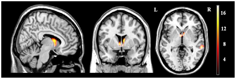
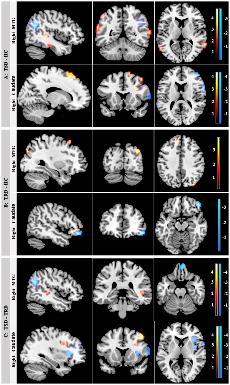
Magnetic resonance imaging (MRI) studies have indicated that the structure deficits and resting-state functional connectivity (FC) imbalances in cortico-limbic circuitry might underline the pathophysiology of MDD. Using structure and functional MRI, our aim is to investigate gray matter abnormalities in patients with treatment-resistant depression (TRD) and treatment-responsive depression (TSD), and test whether the altered gray matter is associated with altered FC. Voxel-based morphometry was used to investigate the regions with gray matter abnormality and FC analysis was further conducted between each gray matter abnormal region and the remaining voxels in the brain. Using one-way analysis of variance, we found significant gray matter abnormalities in the right middle temporal cortex (MTG) and bilateral caudate among the TRD, TSD and healthy controls. For the FC of the right MTG, we found that both the patients with TRD and TSD showed altered connectivity mainly in the default-mode network (DMN). For the FC of the right caudate, both patient groups showed altered connectivity in the frontal regions. Our results revealed the gray matter reduction of right MTG and bilateral caudate, and disrupted functional connection to widely distributed circuitry in DMN and frontal regions, respectively. These results suggest that the abnormal DMN and reward circuit activity might be biomarkers of depression trait.
磁共振成像(MRI)研究表明,皮质边缘回路的结构缺陷和静息状态功能连接(FC)失衡可能是 MDD 病理生理学的基础。本研究采用结构和功能 MRI,旨在探讨治疗抵抗性抑郁症(TRD)和治疗反应性抑郁症(TSD)患者的灰质异常,并检验异常灰质与改变的 FC 是否相关。我们采用基于体素的形态学方法来研究灰质异常区域,进一步在每个灰质异常区域与大脑中其余体素之间进行 FC 分析。通过单因素方差分析,我们发现 TRD、TSD 和健康对照组患者的右侧颞中回(MTG)和双侧尾状核灰质异常。对于右侧 MTG 的 FC,我们发现 TRD 和 TSD 患者的连接均发生改变,主要是在默认模式网络(DMN)中。对于右侧尾状核的 FC,两组患者的额叶区域的连接均发生改变。我们的结果揭示了右侧 MTG 和双侧尾状核的灰质减少,以及与 DMN 和额叶区域广泛分布的回路的功能连接中断,这表明异常的 DMN 和奖励回路活动可能是抑郁特征的生物标志物。