Núcleo de Cognição e Sistemas Complexos, Centro de Matemática, Computação e Cognição, Universidade Federal do ABC, Santo André, São Paulo, Brazil.
PLoS One. 2012;7(9):e45449. doi: 10.1371/journal.pone.0045449. Epub 2012 Sep 20.
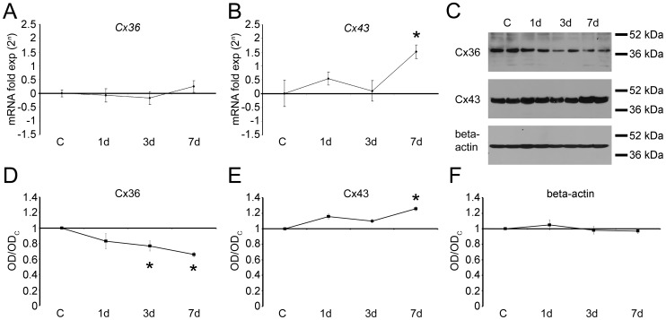
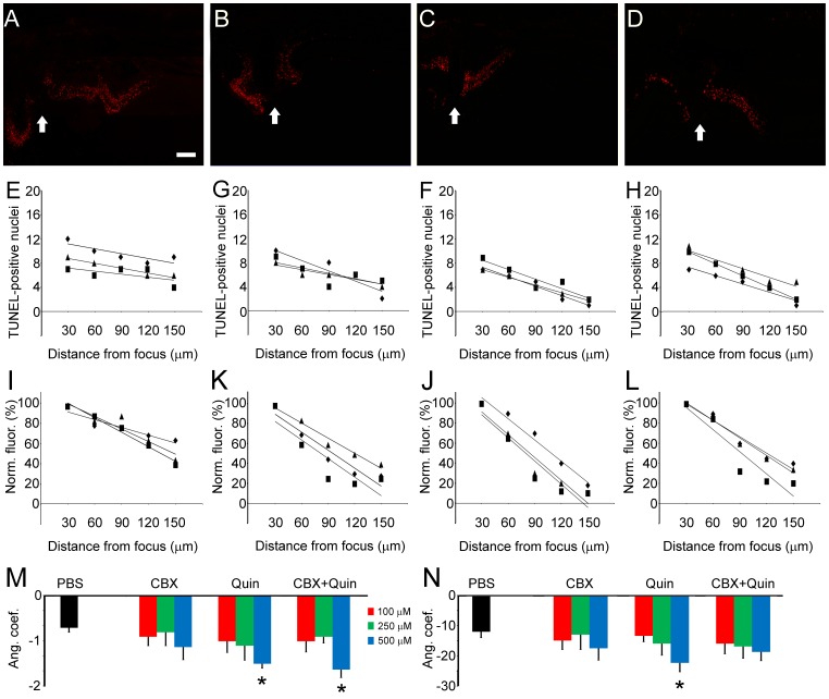
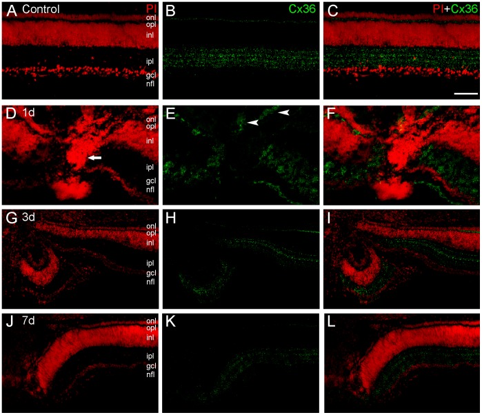
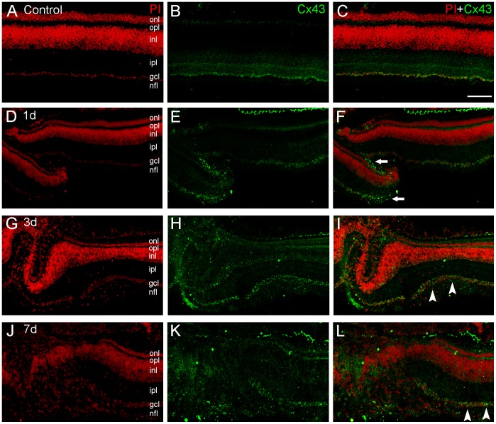
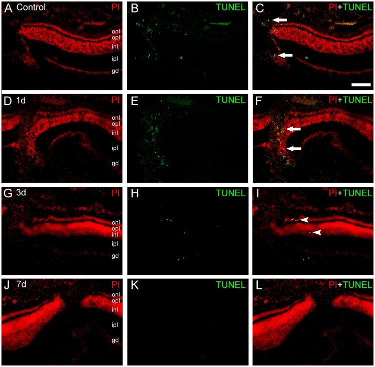
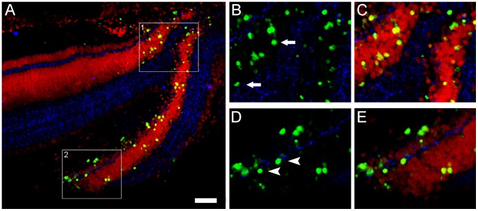
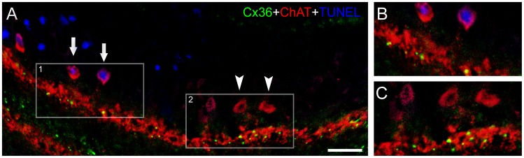
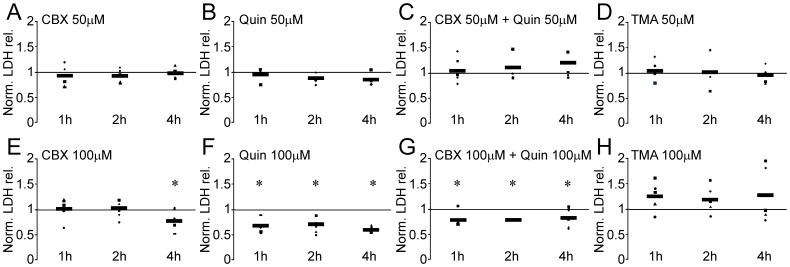
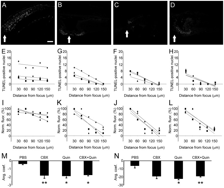
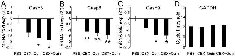
Accruing evidence indicates that connexin (Cx) channels in the gap junctions (GJ) are involved in neurodegeneration after injury. However, studies using KO animal models endowed apparently contradictory results in relation to the role of coupling in neuroprotection. We analyzed the role of Cx-mediated communication in a focal lesion induced by mechanical trauma of the retina, a model that allows spatial and temporal definition of the lesion with high reproducibility, permitting visualization of the focus, penumbra and adjacent areas. Cx36 and Cx43 exhibited distinct gene expression and protein levels throughout the neurodegeneration progress. Cx36 was observed close to TUNEL-positive nuclei, revealing the presence of this protein surrounding apoptotic cells. The functional role of cell coupling was assessed employing GJ blockers and openers combined with lactate dehydrogenase (LDH) assay, a direct method for evaluating cell death/viability. Carbenoxolone (CBX), a broad-spectrum GJ blocker, reduced LDH release after 4 hours, whereas quinine, a Cx36-channel specific blocker, decreased LDH release as early as 1 hour after lesion. Furthermore, analysis of dying cell distribution confirmed that the use of GJ blockers reduced apoptosis spread. Accordingly, blockade of GJ communication during neurodegeneration with quinine, but not CBX, caused downregulation of initial and effector caspases. To summarize, we observed specific changes in Cx gene expression and protein distribution during the progress of retinal degeneration, indicating the participation of these elements in acute neurodegeneration processes. More importantly, our results revealed that direct control of GJ channels permeability may take part in reliable neuroprotection strategies aimed to rapid, fast treatment of mechanical trauma in the retina.
越来越多的证据表明,缝隙连接 (GJ) 中的连接蛋白 (Cx) 通道参与了损伤后的神经退行性变。然而,使用 KO 动物模型的研究在偶联在神经保护中的作用方面得出了明显矛盾的结果。我们分析了 Cx 介导的通讯在机械性视网膜损伤引起的局灶性病变中的作用,该模型允许对病变进行空间和时间的定义,具有高度的可重复性,允许对焦点、半影和相邻区域进行可视化。Cx36 和 Cx43 在整个神经退行性变过程中表现出不同的基因表达和蛋白水平。Cx36 被观察到靠近 TUNEL 阳性核,表明这种蛋白存在于围绕凋亡细胞的周围。通过使用 GJ 阻滞剂和开放剂结合乳酸脱氢酶 (LDH) 测定,评估细胞偶联的功能作用,LDH 测定是一种直接评估细胞死亡/活力的方法。广泛的 GJ 阻滞剂 carbenoxolone (CBX) 减少了 4 小时后的 LDH 释放,而 Cx36 通道特异性阻滞剂 quinine 在损伤后 1 小时就减少了 LDH 释放。此外,对死亡细胞分布的分析证实,使用 GJ 阻滞剂减少了细胞凋亡的扩散。因此,使用 quinine 而不是 CBX 阻断神经退行性变过程中的 GJ 通讯导致初始和效应半胱天冬酶下调。总之,我们观察到 Cx 基因表达和蛋白分布在视网膜变性过程中的特定变化,表明这些元素参与了急性神经退行性变过程。更重要的是,我们的结果表明,直接控制 GJ 通道通透性可能参与可靠的神经保护策略,旨在快速、快速治疗视网膜的机械性创伤。