Department of Neuroscience, Clinical Neurophysiology, Uppsala University, Sweden.
PLoS One. 2012;7(9):e45923. doi: 10.1371/journal.pone.0045923. Epub 2012 Sep 20.
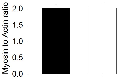
Many mutations in the skeletal muscle α-actin gene (ACTA1) lead to muscle weakness and nemaline myopathy. Despite increasing clinical and scientific interest, the molecular and cellular pathogenesis of weakness remains unclear. Therefore, in the present study, we aimed at unraveling these mechanisms using muscles from a transgenic mouse model of nemaline myopathy expressing the ACTA1 Asp286Gly mutation. We recorded and analyzed the mechanics of membrane-permeabilized single muscle fibers. We also performed molecular energy state computations in the presence or absence of Asp286Gly. Results demonstrated that during contraction, the Asp286Gly acts as a "poison-protein" and according to the computational analysis it modifies the actin-actin interface. This phenomenon is likely to prevent proper myosin cross-bridge binding, limiting the fraction of actomyosin interactions in the strong binding state. At the cell level, this decreases the force-generating capacity, and, overall, induces muscle weakness. To counterbalance such negative events, future potential therapeutic strategies may focus on the inappropriate actin-actin interface or myosin binding.
许多骨骼肌α-肌动蛋白基因 (ACTA1) 的突变导致肌肉无力和杆状体肌病。尽管临床和科学兴趣日益增加,但肌无力的分子和细胞发病机制仍不清楚。因此,在本研究中,我们旨在使用表达 ACTA1 Asp286Gly 突变的杆状体肌病转基因小鼠模型的肌肉来揭示这些机制。我们记录和分析了透膜单根肌纤维的力学特性。我们还在存在或不存在 Asp286Gly 的情况下进行了分子能量状态计算。结果表明,在收缩过程中,Asp286Gly 充当“毒蛋白”,根据计算分析,它改变了肌动蛋白-肌动蛋白界面。这种现象可能会阻止肌球蛋白横桥的正确结合,限制强结合状态下肌动球蛋白相互作用的分数。在细胞水平上,这会降低产生力的能力,并且总体上会导致肌肉无力。为了抵消这种负面影响,未来的潜在治疗策略可能集中在不合适的肌动蛋白-肌动蛋白界面或肌球蛋白结合上。