Centre for Education and Research on Ageing, University of Sydney, Australia.
PLoS One. 2012;7(9):e46134. doi: 10.1371/journal.pone.0046134. Epub 2012 Sep 24.
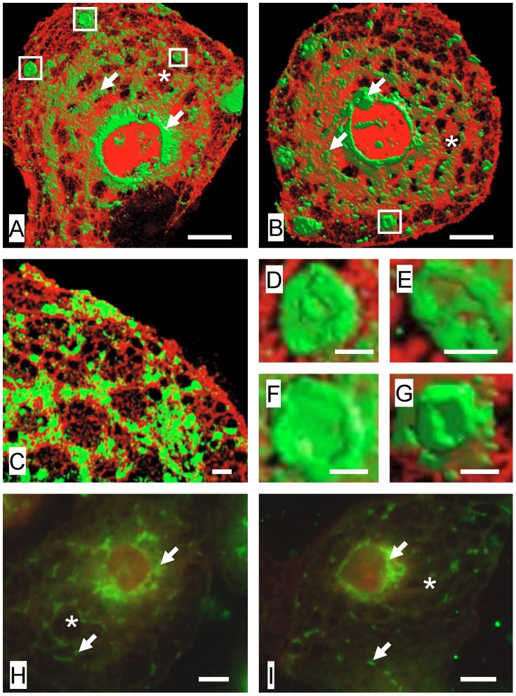
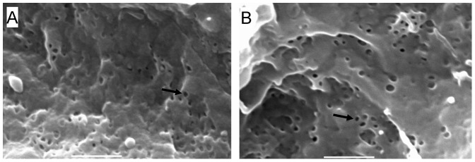
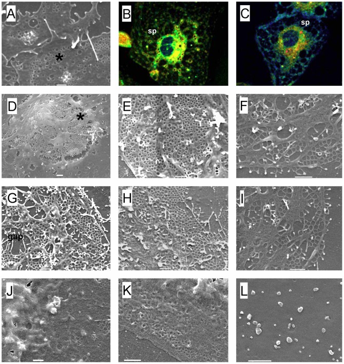
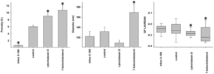
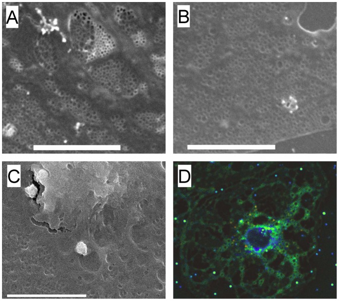
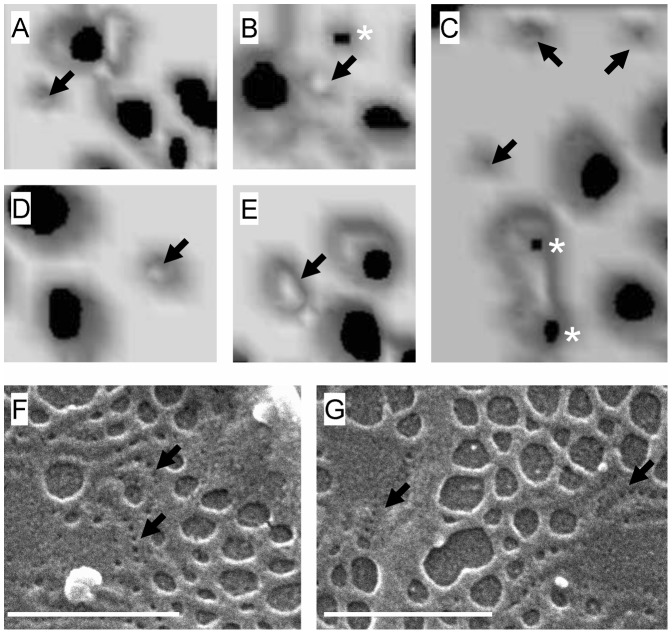
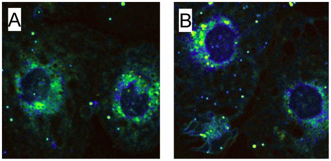
Fenestrations are transcellular pores in endothelial cells that facilitate transfer of substrates between blood and the extravascular compartment. In order to understand the regulation and formation of fenestrations, the relationship between membrane rafts and fenestrations was investigated in liver sinusoidal endothelial cells where fenestrations are grouped into sieve plates. Three dimensional structured illumination microscopy, scanning electron microscopy, internal reflectance fluorescence microscopy and two-photon fluorescence microscopy were used to study liver sinusoidal endothelial cells isolated from mice. There was an inverse distribution between sieve plates and membrane rafts visualized by structured illumination microscopy and the fluorescent raft stain, Bodipy FL C5 ganglioside GM1. 7-ketocholesterol and/or cytochalasin D increased both fenestrations and lipid-disordered membrane, while Triton X-100 decreased both fenestrations and lipid-disordered membrane. The effects of cytochalasin D on fenestrations were abrogated by co-administration of Triton X-100, suggesting that actin disruption increases fenestrations by its effects on membrane rafts. Vascular endothelial growth factor (VEGF) depleted lipid-ordered membrane and increased fenestrations. The results are consistent with a sieve-raft interaction, where fenestrations form in non-raft lipid-disordered regions of endothelial cells once the membrane-stabilizing effects of actin cytoskeleton and membrane rafts are diminished.
窗孔是内皮细胞的细胞间孔,促进血液和血管外腔室之间的底物转移。为了了解窗孔的调节和形成,研究了肝窦内皮细胞中膜筏与窗孔的关系,肝窦内皮细胞中的窗孔被分组为筛板。使用三维结构照明显微镜、扫描电子显微镜、内反射荧光显微镜和双光子荧光显微镜研究从小鼠中分离的肝窦内皮细胞。结构照明显微镜和荧光筏染色剂 Bodipy FL C5 神经节苷脂 GM1 显示,筛板与膜筏之间呈负分布。7-酮胆固醇和/或细胞松弛素 D 增加了窗孔和脂质无序膜,而 Triton X-100 降低了窗孔和脂质无序膜。Triton X-100 共给药可消除细胞松弛素 D 对窗孔的作用,表明肌动蛋白破坏通过其对膜筏的作用增加了窗孔。血管内皮生长因子 (VEGF) 耗尽了有序脂质膜并增加了窗孔。结果与筛-筏相互作用一致,其中一旦肌动蛋白细胞骨架和膜筏的膜稳定作用减弱,窗孔就在内皮细胞的非筏脂质无序区域形成。