Münevveroğlu A P, Aydın K C
Pedodonti Ana Bilim Dalı, Dişhekimliği Fakültesi, İstanbul Medipol Üniversitesi, 34083 Fatih, Turkey.
Case Rep Dent. 2012;2012:654839. doi: 10.1155/2012/654839. Epub 2012 Sep 23.
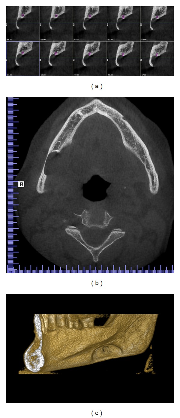
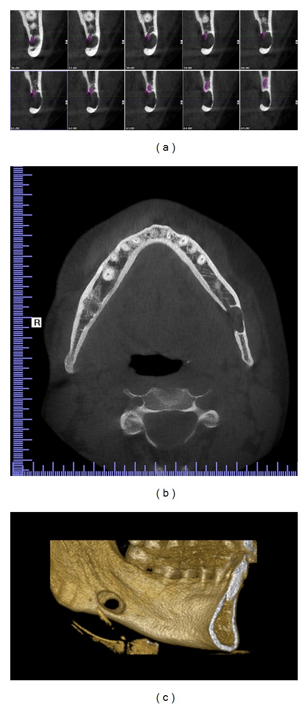
Stafne bone defects are asymptomatic lingual bone depressions of the lower jaw. In 1942, Stafne described for the first time 35 asymptomatic, radiolucent cavities, unilaterally located in the posterior region of the mandible, between the mandibular angle and the third molar, below the inferior dental canal and slightly above the basis mandibulae. In this study, the clinical and radiological characteristics of 2 cases of Stafne bone defects were described. Orthopantomograph and CBCT were used for diagnosing the defects. The bone defects of two patients in this study were asymptomatic and any other bone lesions, such as cysts and tumors, were excluded because no signs of inflammatory or tumoral changes were evident Therefore, surgery was not considered and the patients were followed for 1 year. Stafne bone defect was an incidental finding, presenting no evolutionary changes, and as such conservatory therapy based on periodic controls was indicated. Currently, complementary techniques such as CT are sufficient to establish a certain diagnosis.
斯达姆骨缺损是下颌骨无症状的舌侧骨凹陷。1942年,斯达姆首次描述了35个无症状的放射性透亮腔,这些腔单侧位于下颌骨后部,在下颌角与第三磨牙之间,在下颌管下方且略高于下颌骨基部。在本研究中,描述了2例斯达姆骨缺损的临床和放射学特征。使用全景曲面断层片和锥形束计算机断层扫描(CBCT)来诊断这些缺损。本研究中的两名患者的骨缺损均无症状,且排除了任何其他骨病变,如囊肿和肿瘤,因为没有明显的炎症或肿瘤变化迹象。因此,未考虑手术,对患者进行了1年的随访。斯达姆骨缺损是偶然发现的,没有出现进展性变化,因此建议采用基于定期检查的保守治疗。目前,诸如CT等辅助技术足以做出明确诊断。