• 文献检索
  • 文档翻译
  • 深度研究
  • 学术资讯
  • Suppr Zotero 插件Zotero 插件
  • 邀请有礼
  • 套餐&价格
  • 历史记录
应用&插件
Suppr Zotero 插件Zotero 插件浏览器插件Mac 客户端Windows 客户端微信小程序
定价
高级版会员购买积分包购买API积分包
服务
文献检索文档翻译深度研究API 文档MCP 服务
关于我们
关于 Suppr公司介绍联系我们用户协议隐私条款
关注我们

Suppr 超能文献

核心技术专利:CN118964589B侵权必究
粤ICP备2023148730 号-1Suppr @ 2026

文献检索

告别复杂PubMed语法,用中文像聊天一样搜索,搜遍4000万医学文献。AI智能推荐,让科研检索更轻松。

立即免费搜索

文件翻译

保留排版,准确专业,支持PDF/Word/PPT等文件格式,支持 12+语言互译。

免费翻译文档

深度研究

AI帮你快速写综述,25分钟生成高质量综述,智能提取关键信息,辅助科研写作。

立即免费体验

伴有颊侧皮质骨扩张的斯塔夫内骨腔:病例报告

Stafne's Defect with Buccal Cortical Expansion: A Case Report.

作者信息

Flores Campos Paulo Sérgio, Oliveira José Aloysio Carvalho, Dantas Janaina Araújo, de Melo Daniela Pita, Pena Nilson, Santos Luís Antônio Nogueira, Crusoé-Rebello Iêda Margarida Rocha

机构信息

Department of Radiology, School of Dentistry, Federal University of Bahia, 40110-150 Bahia, Brazil.

出版信息

Int J Dent. 2010;2010:515931. doi: 10.1155/2010/515931. Epub 2010 May 4.

DOI:10.1155/2010/515931
PMID:20454575
原文链接:https://pmc.ncbi.nlm.nih.gov/articles/PMC2864448/
Abstract

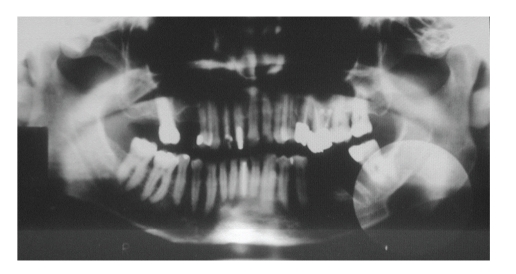
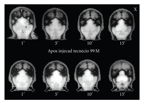
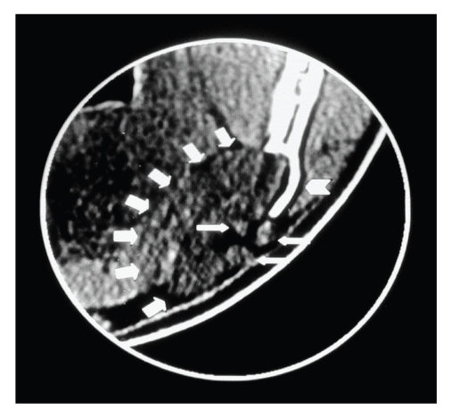
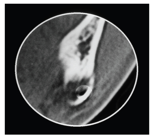
A rare case of Stafne's bone cavity, type III-G, is reported in a 49-year-old male patient who had been referred to a private clinic for a routine evaluation. The final diagnosis was based on computed tomography. Scintigraphy played a fundamental role in determining the most likely etiology.

摘要

报告了一例罕见的III - G型斯塔夫内骨腔病例,患者为一名49岁男性,因常规评估被转诊至一家私人诊所。最终诊断基于计算机断层扫描。骨闪烁显像在确定最可能的病因方面发挥了重要作用。

https://cdn.ncbi.nlm.nih.gov/pmc/blobs/eb10/2864448/c4d7a9152dbf/IJD2010-515931.004.jpg
https://cdn.ncbi.nlm.nih.gov/pmc/blobs/eb10/2864448/2dda26a37590/IJD2010-515931.001.jpg
https://cdn.ncbi.nlm.nih.gov/pmc/blobs/eb10/2864448/010ecc68f8fa/IJD2010-515931.002.jpg
https://cdn.ncbi.nlm.nih.gov/pmc/blobs/eb10/2864448/1f75e8e66559/IJD2010-515931.003.jpg
https://cdn.ncbi.nlm.nih.gov/pmc/blobs/eb10/2864448/c4d7a9152dbf/IJD2010-515931.004.jpg

相似文献

1
Stafne's Defect with Buccal Cortical Expansion: A Case Report.伴有颊侧皮质骨扩张的斯塔夫内骨腔:病例报告
Int J Dent. 2010;2010:515931. doi: 10.1155/2010/515931. Epub 2010 May 4.
2
Stafne's Bone Defect Correlated with Submandibular Glands: A Case Report and CBCT and MRI Analysis.与下颌下腺相关的斯塔夫内骨缺损:一例报告及CBCT和MRI分析
Case Rep Dent. 2024 Feb 6;2024:1173783. doi: 10.1155/2024/1173783. eCollection 2024.
3
Stafne's bone defect with bicortical perforation: a need for modified classification system.Stafne 氏骨缺损伴皮质穿孔:需要改良的分类系统。
Oral Radiol. 2021 Jan;37(1):130-136. doi: 10.1007/s11282-020-00457-8. Epub 2020 Jun 22.
4
Stafne's bone defect: a case report and review of literatures.斯塔夫内骨缺损:一例病例报告及文献综述
Ann Transl Med. 2019 Aug;7(16):399. doi: 10.21037/atm.2019.07.73.
5
Stafne's bone defects from Spain: report of four cases and brief archaeological literature review.西班牙的斯塔夫内骨缺损:4例报告及考古文献简要综述
Int J Paleopathol. 2020 Dec;31:46-52. doi: 10.1016/j.ijpp.2020.08.001. Epub 2020 Sep 16.
6
Stafne's Bone Cavity: A Diagnostic Challenge.斯塔夫内骨腔:一项诊断挑战。
J Clin Diagn Res. 2015 Nov;9(11):ZD16-9. doi: 10.7860/JCDR/2015/14273.6772. Epub 2015 Nov 1.
7
An unilateral basal bone defect of the mandible occupied by fatty tissue: Stafne's cavity.单侧下颌骨基底骨缺损被脂肪组织占据:Stafne 腔。
In Vivo. 2012 Nov-Dec;26(6):1045-8.
8
Stafne's bone cavity: An unusual case with involvement of the buccal and lingual mandibular plates.斯塔夫内骨腔:累及下颌颊侧和舌侧骨板的罕见病例。
J Clin Exp Dent. 2014 Feb 1;6(1):e96-9. doi: 10.4317/jced.51229. eCollection 2014 Feb.
9
Mandibular ramus-related Stafne's bone cavity.下颌支相关的斯塔夫内骨腔。
Dentomaxillofac Radiol. 2004 Jan;33(1):63-6. doi: 10.1259/dmfr/39682286.
10
CT analysis of the Stafne's bone defects of the mandible.下颌骨斯丹氏骨缺损的CT分析
Dentomaxillofac Radiol. 2006 Mar;35(2):95-102. doi: 10.1259/dmfr/71115878.

引用本文的文献

1
Radiological features of Stafne mandibular bone cavity in panoramic image and cone beam computed tomography.全景图像和锥形束计算机断层扫描中斯陶德下颌骨腔的放射学特征
Maxillofac Plast Reconstr Surg. 2024 Mar 19;46(1):9. doi: 10.1186/s40902-024-00415-y.
2
Evaluating Static Bone Cysts (SBCs) Through Long-Term Volumetric Analysis Using CBCT: A Study with 6-Month Follow-Up.使用锥形束计算机断层扫描(CBCT)通过长期容积分析评估静态骨囊肿(SBCs):一项为期6个月随访的研究。
J Clin Exp Dent. 2024 Feb 1;16(2):e104-e110. doi: 10.4317/jced.61099. eCollection 2024 Feb.
3
Imaging features of Stafne bone defects on computed tomography: An assessment of 40 cases.

本文引用的文献

1
CT analysis of the Stafne's bone defects of the mandible.下颌骨斯丹氏骨缺损的CT分析
Dentomaxillofac Radiol. 2006 Mar;35(2):95-102. doi: 10.1259/dmfr/71115878.
2
Mandibular ramus-related Stafne's bone cavity.下颌支相关的斯塔夫内骨腔。
Dentomaxillofac Radiol. 2004 Jan;33(1):63-6. doi: 10.1259/dmfr/39682286.
3
Etiology of static bone defects of the mandible.下颌骨静止性骨缺损的病因
计算机断层扫描下斯陶芬骨缺损的影像学特征:40例病例评估
Imaging Sci Dent. 2021 Mar;51(1):81-86. doi: 10.5624/isd.20200253. Epub 2021 Jan 29.
4
Stafne's bone defect with bicortical perforation: a need for modified classification system.Stafne 氏骨缺损伴皮质穿孔:需要改良的分类系统。
Oral Radiol. 2021 Jan;37(1):130-136. doi: 10.1007/s11282-020-00457-8. Epub 2020 Jun 22.
5
Stafne's bone cyst revisited and renamed: the benign mandibular concavity.重新审视并更名为 Stafne 氏骨囊肿:良性下颌骨凹陷。
Dentomaxillofac Radiol. 2020 May 1;49(4):20190475. doi: 10.1259/dmfr.20190475. Epub 2020 Feb 3.
6
Stafne bone cavity and cone-beam computed tomography: a report of two cases.斯丹佛骨腔与锥形束计算机断层扫描:两例报告
J Korean Assoc Oral Maxillofac Surg. 2015 Jun;41(3):145-8. doi: 10.5125/jkaoms.2015.41.3.145. Epub 2015 Jun 23.
7
Evaluation of volumetric measurements on CBCT images using stafne bone cavities as an example.以斯丹纳骨腔为例评估锥形束计算机断层扫描(CBCT)图像上的容积测量
Med Oral Patol Oral Cir Bucal. 2015 Sep 1;20(5):e580-6. doi: 10.4317/medoral.20633.
8
Stafne bone defect: report of two cases.斯塔夫内骨缺损:两例报告。
Case Rep Dent. 2012;2012:654839. doi: 10.1155/2012/654839. Epub 2012 Sep 23.
J Oral Surg Anesth Hosp Dent Serv. 1960 Jan;18:16-20.
4
Evaluation of static bone cavities with CT and MRI.利用CT和MRI评估静态骨腔。
Dentomaxillofac Radiol. 2003 Jan;32(1):2-7. doi: 10.1259/dmfr/92201340.
5
Lingual and buccal mandibular bone depressions: a review based on 583 cases from a world-wide literature survey, including 69 new cases from Japan.舌侧和颊侧下颌骨凹陷:基于一项全球文献调查中583例病例的综述,其中包括来自日本的69例新病例。
Dentomaxillofac Radiol. 2002 Sep;31(5):281-90. doi: 10.1038/sj.dmfr.4600718.
6
Stafne's bone cavity. Classification based on outline and content determined by computed tomography.斯塔夫内骨腔。基于计算机断层扫描所确定的轮廓和内容的分类。
Oral Surg Oral Med Oral Pathol. 1993 Sep;76(3):375-80. doi: 10.1016/0030-4220(93)90271-5.
7
Developmental bone defects of the mandible.
Oral Surg Oral Med Oral Pathol. 1967 Oct;24(4):488-90. doi: 10.1016/0030-4220(67)90423-9.