Martin Chris, Zheng Ying, Sibson Nicola R, Mayhew John E W, Berwick Jason
Department of Psychology, University of Sheffield, Western Bank, Sheffield, S10 2TP, UK.
Department of Psychology, University of Sheffield, Western Bank, Sheffield, S10 2TP, UK.
Neuroimage. 2013 Feb 1;66:1-8. doi: 10.1016/j.neuroimage.2012.10.006. Epub 2012 Oct 11.
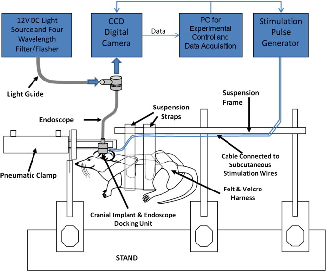
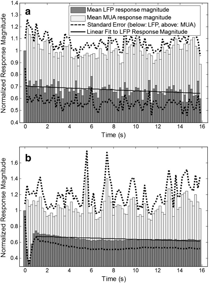
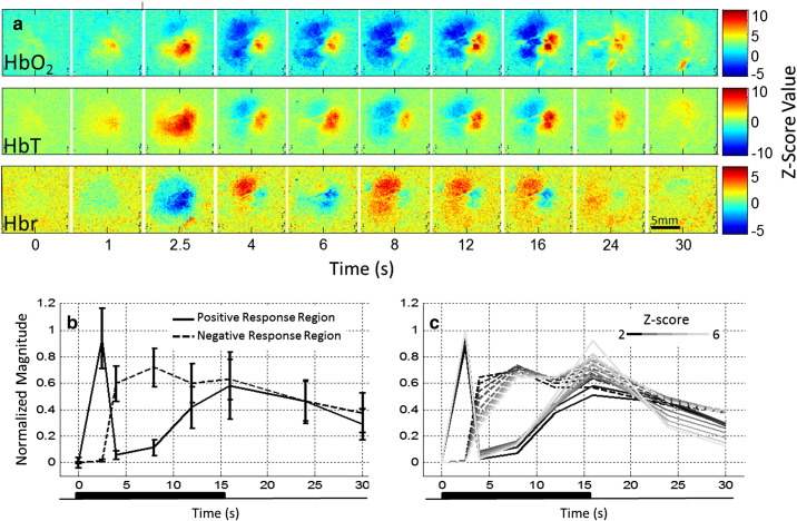
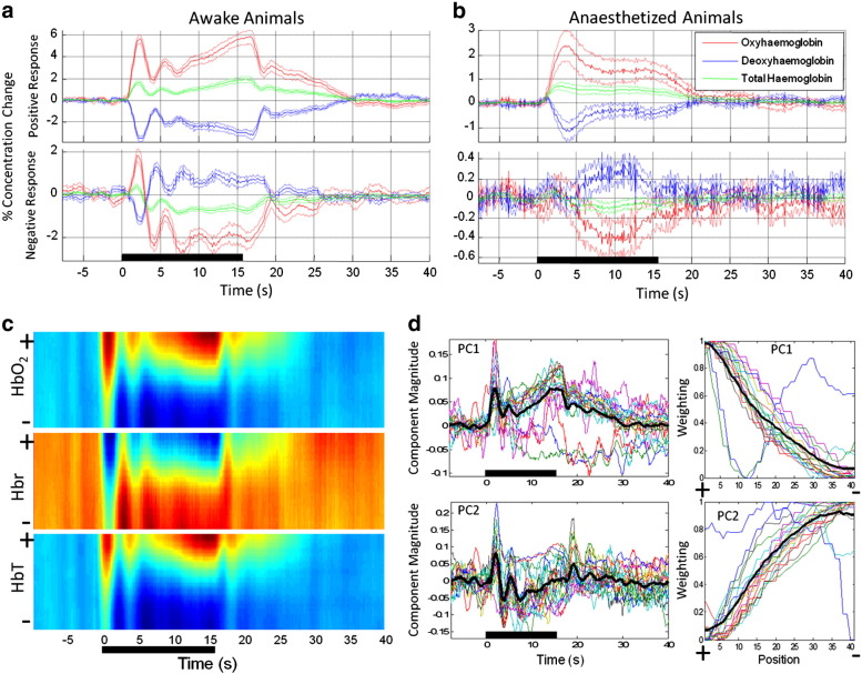
Detailed understanding of the haemodynamic changes that underlie non-invasive neuroimaging techniques such as blood oxygen level dependent functional magnetic resonance imaging is essential if we are to continue to extend the use of these methods for understanding brain function and dysfunction. The use of animal and in particular rodent research models has been central to these endeavours as they allow in-vivo experimental techniques that provide measurements of the haemodynamic response function at high temporal and spatial resolution. A limitation of most of this research is the use of anaesthetic agents which may disrupt or mask important features of neurovascular coupling or the haemodynamic response function. In this study we therefore measured spatiotemporal cortical haemodynamic responses to somatosensory stimulation in awake rats using optical imaging spectroscopy. Trained, restrained animals received non-noxious stimulation of the whisker pad via chronically implanted stimulating microwires whilst optical recordings were made from the contralateral somatosensory cortex through a thin cranial window. The responses we measure from un-anaesthetised animals are substantially different from those reported in previous studies which have used anaesthetised animals. These differences include biphasic response regions (initial increases in blood volume and oxygenation followed by subsequent decreases) as well as oscillations in the response time series of awake animals. These haemodynamic response features do not reflect concomitant changes in the underlying neuronal activity and therefore reflect neurovascular or cerebrovascular processes. These hitherto unreported hyperemic response dynamics may have important implications for the use of anaesthetised animal models for research into the haemodynamic response function.
如果我们要继续扩大使用这些方法来理解脑功能和功能障碍,那么深入了解诸如血氧水平依赖性功能磁共振成像等非侵入性神经成像技术背后的血流动力学变化至关重要。动物尤其是啮齿动物研究模型的使用一直是这些努力的核心,因为它们允许采用体内实验技术,以高时间和空间分辨率提供血流动力学响应函数的测量。这项研究的局限性在于,大多数研究使用了麻醉剂,这可能会干扰或掩盖神经血管耦合或血流动力学响应函数的重要特征。因此,在本研究中,我们使用光学成像光谱法测量了清醒大鼠体感刺激的时空皮质血流动力学响应。经过训练和约束的动物通过长期植入的刺激微丝接受对须垫的无害刺激,同时通过薄颅窗从对侧体感皮层进行光学记录。我们从未麻醉动物身上测量到的反应与先前使用麻醉动物的研究报告有很大不同。这些差异包括双相反应区域(血容量和氧合先增加后减少)以及清醒动物反应时间序列中的振荡。这些血流动力学反应特征并不反映潜在神经元活动的伴随变化,因此反映了神经血管或脑血管过程。这些迄今未报告的充血反应动态可能对使用麻醉动物模型研究血流动力学响应函数具有重要意义。