Lee Jung Uee, Sul Hae Joung, Son Ji Woong
Department of Pathology, St. Mary's Hospital, The Catholic University of Korea School of Medicine, Daejeon, Korea.
Tuberc Respir Dis (Seoul). 2012 Jul;73(1):11-21. doi: 10.4046/trd.2012.73.1.11. Epub 2012 Jul 31.
While qualitative analysis of methylation has been reviewed, the quantitative analysis of methylation has rarely been studied. We evaluated the methylation status of CDKN2A, RARβ, and RASSF1A promoter regions in non-small cell lung carcinomas (NSCLCs) by using pyrosequencing. Then, we evaluated the association between methylation at the promoter regions of these tumor suppressor genes and the clinicopathological parameters of the NSCLCs.
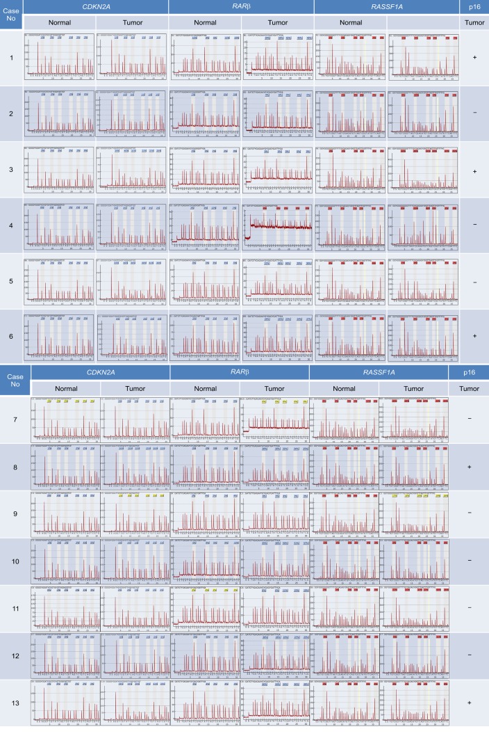
We collected tumor tissues from a total of 53 patients with NSCLCs and analyzed the methylation level of the CDKN2A, RARβ, and RASSF1A promoter regions by using pyrosequencing. In addition, we investigated the correlation between the hypermethylation of CDKN2A and the loss of p16(INK4A) immunoexpression.
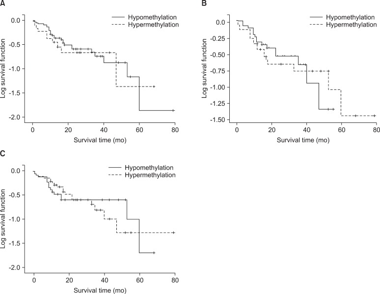
Hypermethylation of CDKN2A, RARβ, and RASSF1A promoter regions were 16 (30.2%), 22 (41.5%), and 21 tumors (39.6%), respectively. The incidence of hypermethylation at the CDKN2A promoter in the tumors was higher in undifferentiated large cell carcinomas than in other subtypes (p=0.002). Hyperrmethylation of CDKN2A was significantly associated with p16(INK4A) immunoexpression loss (p=0.045). With regard to the clinicopathological characteristics of NSCLC, certain histopathological subtypes were found to be strongly associated with the loss of p16(INK4A) immunoexpression (p=0.016). Squamous cell carcinoma and undifferentiated large cell carcinoma showed p16(INK4A) immunoexpression loss more frequently. The Kaplan-Meier survival curves analysis showed that methylation level and patient survival were barely related to one another.
We quantitatively analyzed the promoter methylation status by using pyrosequencing. We showed a significant correlation between CDKN2A hypermethylation and p16(INK4A) immunoexpression loss.
虽然已经对甲基化的定性分析进行了综述,但甲基化的定量分析却很少被研究。我们通过焦磷酸测序评估了非小细胞肺癌(NSCLC)中CDKN2A、RARβ和RASSF1A启动子区域的甲基化状态。然后,我们评估了这些肿瘤抑制基因启动子区域的甲基化与NSCLC临床病理参数之间的关联。
我们收集了总共53例NSCLC患者的肿瘤组织,并通过焦磷酸测序分析了CDKN2A、RARβ和RASSF1A启动子区域的甲基化水平。此外,我们研究了CDKN2A的高甲基化与p16(INK4A)免疫表达缺失之间的相关性。
CDKN2A、RARβ和RASSF1A启动子区域的高甲基化分别见于16例(30.2%)、22例(41.5%)和21例肿瘤(39.6%)。未分化大细胞癌中肿瘤CDKN2A启动子的高甲基化发生率高于其他亚型(p = 0.002)。CDKN2A的高甲基化与p16(INK4A)免疫表达缺失显著相关(p = 0.045)。关于NSCLC的临床病理特征,发现某些组织病理学亚型与p16(INK4A)免疫表达缺失密切相关(p = 0.016)。鳞状细胞癌和未分化大细胞癌更频繁地出现p16(INK4A)免疫表达缺失。Kaplan-Meier生存曲线分析表明,甲基化水平与患者生存率几乎没有相关性。
我们通过焦磷酸测序定量分析了启动子甲基化状态。我们显示CDKN2A高甲基化与p16(INK4A)免疫表达缺失之间存在显著相关性。