TransTissue Technologies GmbH, Berlin, Germany.
J Orthop Surg Res. 2012 Nov 9;7:37. doi: 10.1186/1749-799X-7-37.
Scaffold-assisted autologous chondrocyte implantation is an effective clinical procedure for cartilage repair. From the regulatory point of view, the ovine model is one of the suggested large animal models for pre-clinical studies. The aim of our study was to evaluate the in vitro re-differentiation capacity of expanded ovine chondrocytes in biomechanically characterized polyglycolic acid (PGA)/fibrin biomaterials for scaffold-assisted cartilage repair.
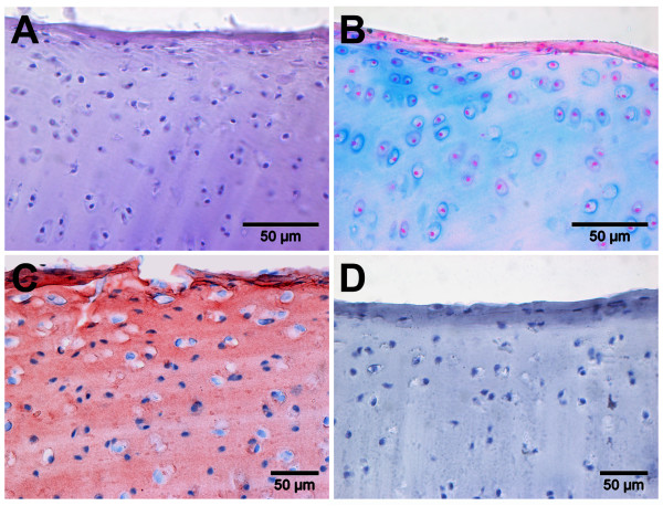
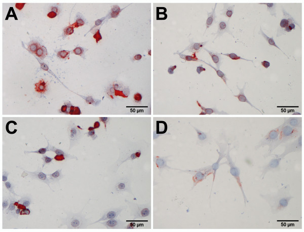
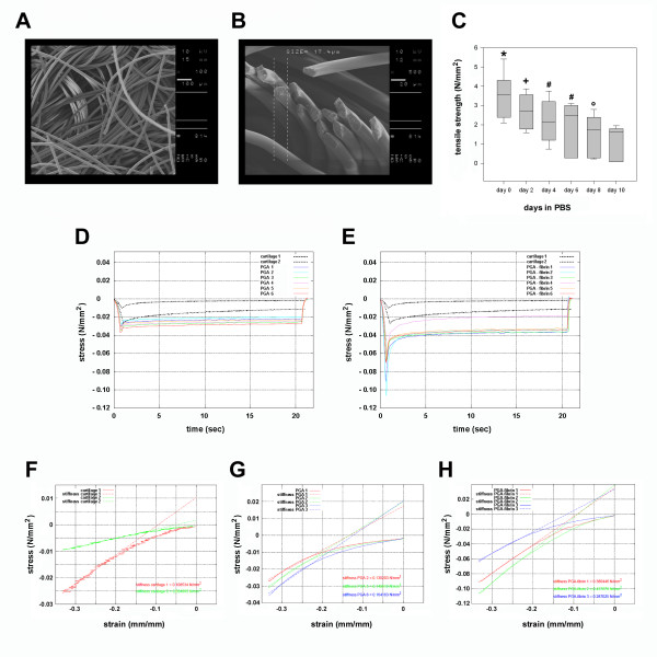
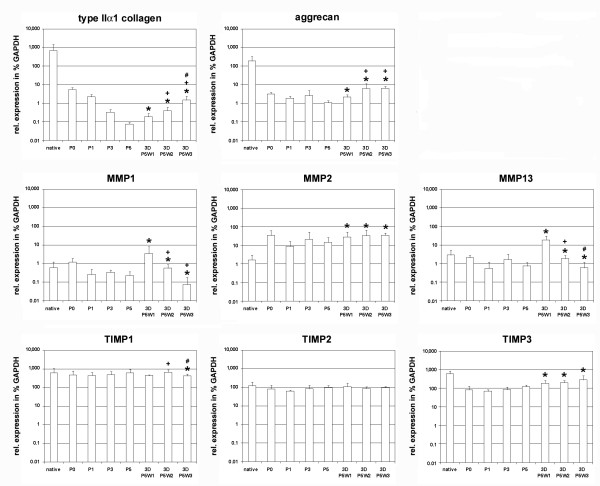
Ovine chondrocytes harvested from adult articular cartilage were expanded in monolayer and re-assembled three-dimensionally in PGA-fibrin scaffolds. De- and re-differentiation of ovine chondrocytes in PGA-fibrin scaffolds was assessed by histological and immuno-histochemical staining as well as by real-time gene expression analysis of typical cartilage marker molecules and the matrix-remodelling enzymes matrix metalloproteinases (MMP) -1, -2 and -13 as well as their inhibitors. PGA scaffolds characteristics including degradation and stiffness were analysed by electron microscopy and biomechanical testing.
Histological, immuno-histochemical and gene expression analysis showed that dedifferentiated chondrocytes re-differentiate in PGA-fibrin scaffolds and form a cartilaginous matrix. Re-differentiation was accompanied by the induction of type II collagen and aggrecan, while MMP expression decreased in prolonged tissue culture. Electron microscopy and biomechanical tests revealed that the non-woven PGA scaffold shows a textile structure with high tensile strength of 3.6 N/mm2 and a stiffness of up to 0.44 N/mm2, when combined with gel-like fibrin.
These data suggest that PGA-fibrin is suited as a mechanically stable support structure for scaffold-assisted chondrocyte grafts, initiating chondrogenic re-differentiation of expanded chondrocytes.
支架辅助自体软骨细胞移植是一种有效的软骨修复临床方法。从监管的角度来看,绵羊模型是临床前研究中建议的大型动物模型之一。我们的研究目的是评估在生物力学特性聚乙二醇酸(PGA)/纤维蛋白生物材料中扩展的绵羊软骨细胞的体外再分化能力,用于支架辅助软骨修复。
从成年关节软骨中收获的绵羊软骨细胞在单层中扩增,并在 PGA-纤维蛋白支架中重新组装成三维结构。通过组织学和免疫组织化学染色以及实时基因表达分析典型的软骨标记分子以及基质重塑酶基质金属蛋白酶(MMP)-1、-2 和 -13 及其抑制剂,评估 PGA-纤维蛋白支架中去分化和再分化的绵羊软骨细胞。通过电子显微镜和生物力学测试分析 PGA 支架的特性,包括降解和刚度。
组织学、免疫组织化学和基因表达分析表明,去分化的软骨细胞在 PGA-纤维蛋白支架中再分化并形成软骨基质。再分化伴随着 II 型胶原和聚集蛋白的诱导,而 MMP 的表达在延长的组织培养中降低。电子显微镜和生物力学测试显示,非织造 PGA 支架与凝胶状纤维蛋白结合时具有纺织结构,拉伸强度为 3.6 N/mm2,刚度高达 0.44 N/mm2。
这些数据表明,PGA-纤维蛋白适合作为支架辅助软骨细胞移植物的机械稳定支撑结构,可启动扩展软骨细胞的软骨生成再分化。