Laboratory of Biochemistry and Cellular Biology (URBC), NARILIS, University of Namur - FUNDP, Belgium.
PLoS One. 2012;7(11):e47519. doi: 10.1371/journal.pone.0047519. Epub 2012 Nov 5.
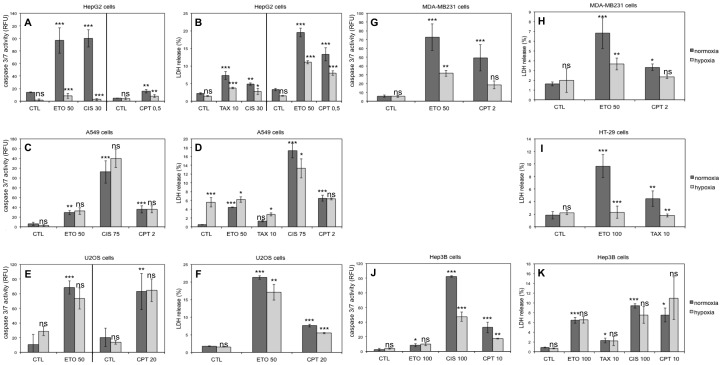
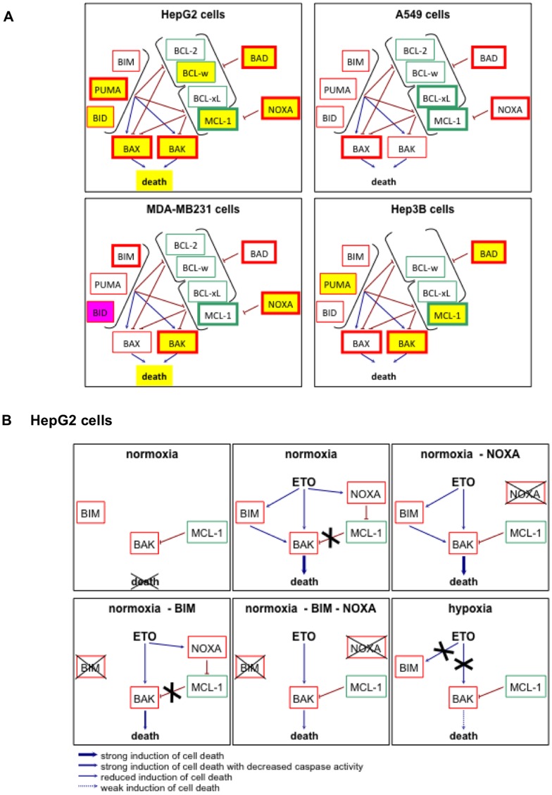
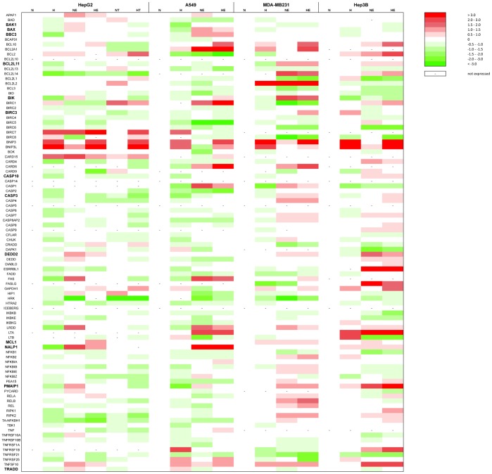
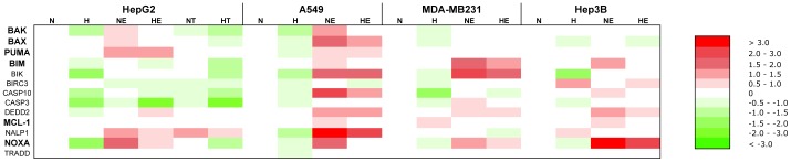
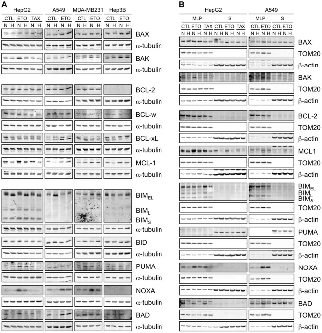
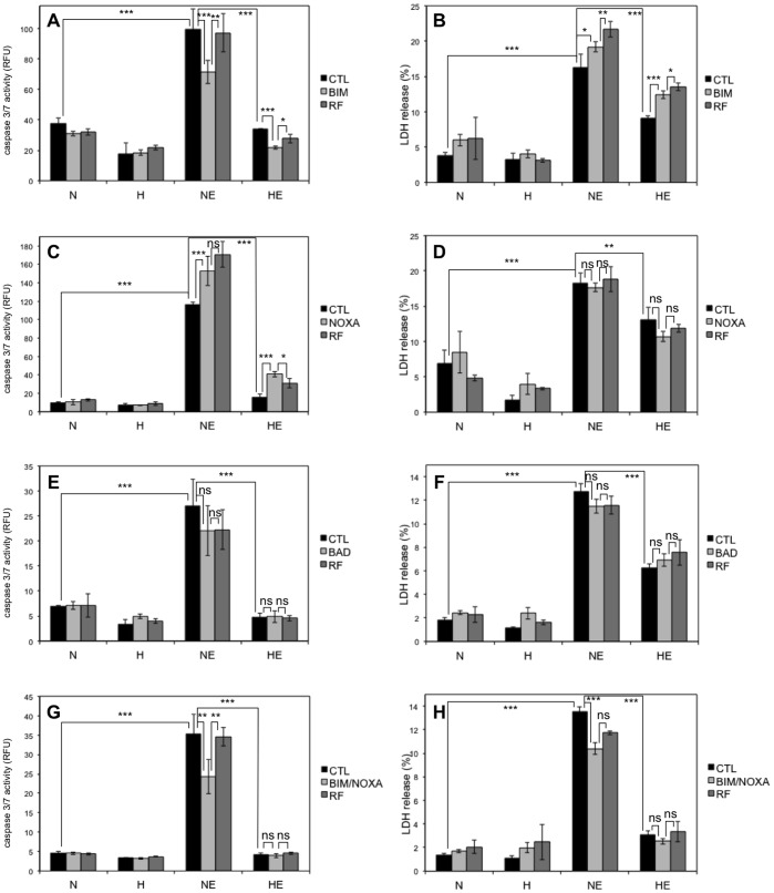
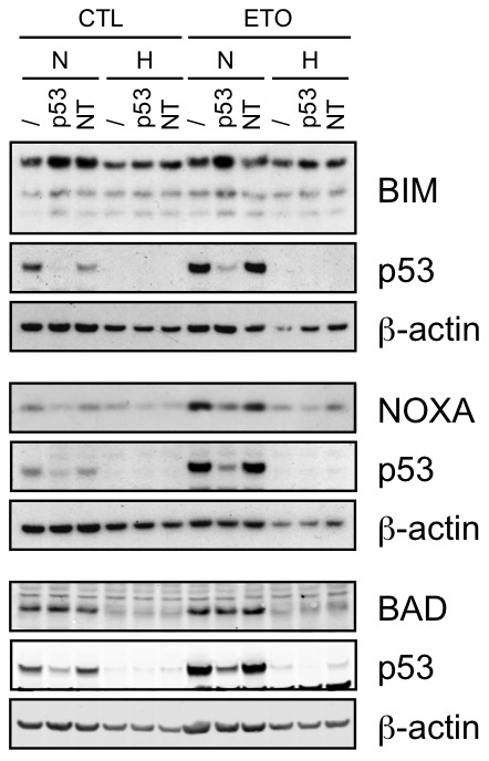
Hypoxia plays an important role in the resistance of tumour cells to chemotherapy. However, the exact mechanisms underlying this process are not well understood. Moreover, according to the cell lines, hypoxia differently influences cell death. The study of the effects of hypoxia on the apoptosis induced by 5 chemotherapeutic drugs in 7 cancer cell types showed that hypoxia generally inhibited the drug-induced apoptosis. In most cases, the effect of hypoxia was the same for all the drugs in one cell type. The expression profile of 93 genes involved in apoptosis as well as the protein level of BCL-2 family proteins were then investigated. In HepG2 cells that are strongly protected against cell death by hypoxia, hypoxia decreased the abundance of nearly all the pro-apoptotic BCL-2 family proteins while none of them are decreased in A549 cells that are not protected against cell death by hypoxia. In HepG2 cells, hypoxia decreased NOXA and BAD abundance and modified the electrophoretic mobility of BIM(EL). BIM and NOXA are important mediators of etoposide-induced cell death in HepG2 cells and the hypoxia-induced modification of these proteins abundance or post-translational modifications partly account for chemoresistance. Finally, the modulation of the abundance and/or of the post-translational modifications of most proteins of the BCL-2 family by hypoxia involves p53-dependent and -independent pathways and is cell type-dependent. A better understanding of these cell-to-cell variations is crucial in order to overcome hypoxia-induced resistance and to ameliorate cancer therapy.
缺氧在肿瘤细胞对化疗的耐药性中起着重要作用。然而,这一过程的确切机制尚不清楚。此外,根据细胞系的不同,缺氧对细胞死亡的影响也不同。研究缺氧对 7 种癌细胞类型中 5 种化疗药物诱导的细胞凋亡的影响表明,缺氧通常抑制药物诱导的细胞凋亡。在大多数情况下,缺氧对一种细胞类型中所有药物的影响是相同的。然后研究了参与凋亡的 93 个基因的表达谱以及 BCL-2 家族蛋白的蛋白质水平。在缺氧强烈保护 HepG2 细胞免受细胞死亡的情况下,缺氧降低了几乎所有促凋亡的 BCL-2 家族蛋白的丰度,而在缺氧不能保护 A549 细胞免受细胞死亡的情况下,它们都没有降低。在 HepG2 细胞中,缺氧降低了 NOXA 和 BAD 的丰度,并改变了 BIM(EL)的电泳迁移率。BIM 和 NOXA 是 HepG2 细胞中依托泊苷诱导细胞死亡的重要介质,这些蛋白质丰度或翻译后修饰的缺氧诱导修饰部分解释了化疗耐药性。最后,缺氧对 BCL-2 家族大多数蛋白质的丰度和/或翻译后修饰的调节涉及 p53 依赖性和非依赖性途径,并且依赖于细胞类型。更好地理解这些细胞间的差异对于克服缺氧诱导的耐药性和改善癌症治疗至关重要。