Sherbrooke Molecular Imaging Center, Department of Nuclear Medicine & Radiobiology, Faculty of Medicine and Health Sciences, Université de Sherbrooke, 3001, 12th Avenue N,, Sherbrooke, Québec, J1H 5N4, Canada.
EJNMMI Res. 2012 Nov 9;2(1):61. doi: 10.1186/2191-219X-2-61.
The estrogen receptor α (ERα) is known to play an important role in the modulation of tumor response to hormone therapy. In this work, the effect of different hormone therapies on tumors having different ERα expression levels was followed up in vivo in a mouse model by PET imaging using 2-deoxy-2-[18F]fluoro-d-glucose (FDG) and [11C]-methionine ([11C]-MET). A new model of MC7-L1 ERα-knockdown (ERαKD) tumor cell lines was designed as a negative estrogen receptor control to follow up the effects of changes in ERα expression on the early metabolic tumor response to different hormone therapies.
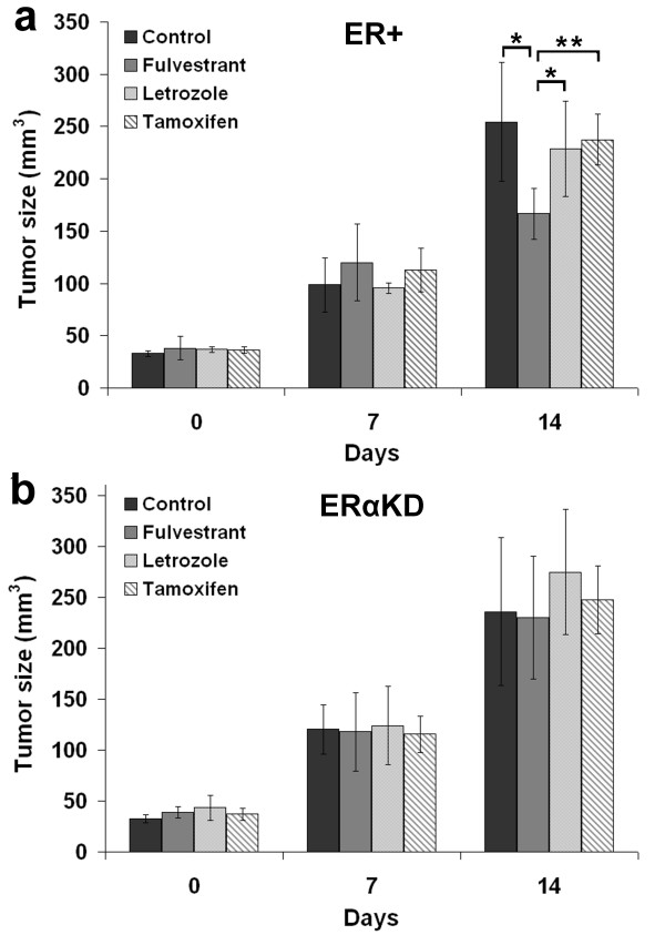
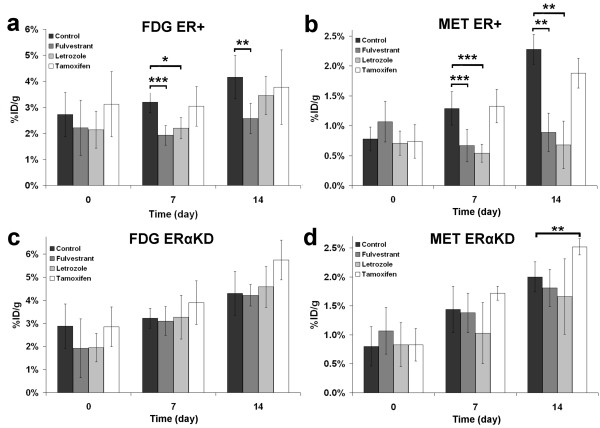
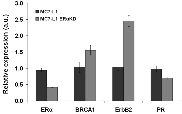
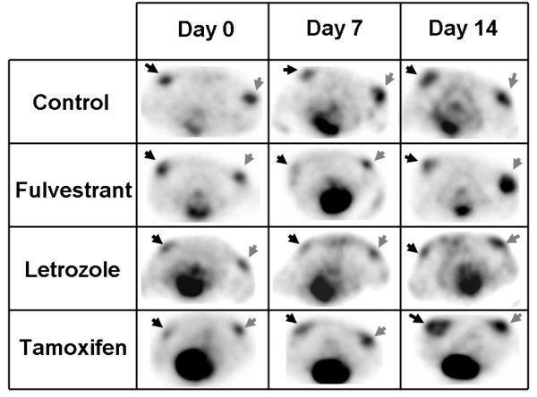
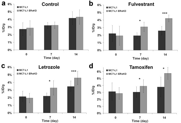
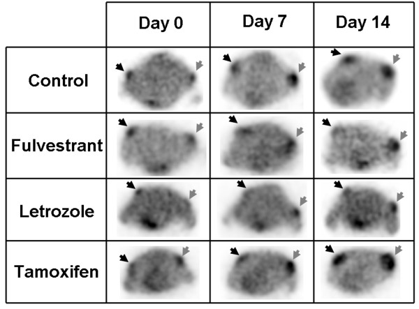
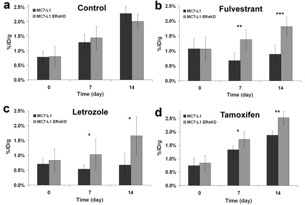
MC7-L1 (ER+) and MC7-L1 ERα-knockdown cell lines were implanted subcutaneously in Balb/c mice and allowed to grow up to 4 mm in diameter. Animals were separated into 4 groups (n = 4 or 5) and treated with a pure antiestrogen (fulvestrant), an aromatase inhibitor (letrozole), a selective estrogen receptor modulator (tamoxifen), or not treated (control). Tumor metabolic activity was assessed by PET imaging with FDG and [11C]-MET at days 0 (before treatment), 7, and 14 after the treatment. Tumor uptake of each radiotracer in %ID/g was measured for each tumor at each time point and compared to tumor growth. Quantitative PCR (qPCR) was performed to verify the expression of breast cancer-related genes (ERα, ErbB2, progesterone receptor (PR), and BRCA1) in each tumor cell lines.
While both ER+ and ERαKD tumors had similar uptake of both radiotracers without treatment, higher uptake values were generally seen in ERαKD tumors after 7 and 14 days of treatment, indicating that ERαKD tumors behave in a similar fashion as hormone-unresponsive tumors. Furthermore, the ERα-specific downregulation induced a slight PR expression decrease and overexpression of BRCA1 and ErbB2.
The results indicate that the proposed ER+/ERαKD tumor-bearing mouse model is suitable to test pure antiestrogen and aromatase inhibitor therapies in vivo in a preclinical setting and could help to elucidate the impact of ERα levels on tumor response to hormone therapy.
雌激素受体 α (ERα) 被认为在调节肿瘤对激素治疗的反应中发挥重要作用。在这项工作中,通过使用 2-脱氧-2-[18F]氟-D-葡萄糖 (FDG) 和 [11C]-蛋氨酸 ([11C]-MET) 的 PET 成像,在一种小鼠模型中对具有不同 ERα 表达水平的肿瘤进行了体内跟踪,以研究不同激素治疗对肿瘤的影响。设计了一种新的 MC7-L1 ERα 敲低 (ERαKD) 肿瘤细胞系模型作为阴性雌激素受体对照,以跟踪 ERα 表达变化对不同激素治疗早期代谢肿瘤反应的影响。
MC7-L1(ER+)和 MC7-L1 ERα 敲低细胞系皮下植入 Balb/c 小鼠,直至肿瘤直径达到 4 毫米。将动物分为 4 组(n=4 或 5),分别用纯抗雌激素(氟维司群)、芳香化酶抑制剂(来曲唑)、选择性雌激素受体调节剂(他莫昔芬)或不治疗(对照组)进行治疗。在治疗前(第 0 天)、治疗后第 7 天和第 14 天,通过 FDG 和 [11C]-MET 的 PET 成像评估肿瘤代谢活性。在每个时间点测量每个肿瘤的每个示踪剂的放射性摄取百分比(%ID/g),并与肿瘤生长进行比较。对每个肿瘤细胞系进行定量 PCR (qPCR) 以验证乳腺癌相关基因(ERα、ErbB2、孕激素受体 (PR) 和 BRCA1)的表达。
在未治疗的情况下,ER+和 ERαKD 肿瘤对两种示踪剂的摄取相似,但在治疗后 7 天和 14 天,ERαKD 肿瘤的摄取值通常更高,表明 ERαKD 肿瘤的行为类似于激素无反应性肿瘤。此外,ERα 的特异性下调导致 PR 表达略有下降,BRCA1 和 ErbB2 过表达。
结果表明,所提出的 ER+/ERαKD 荷瘤小鼠模型适合在临床前环境中体内测试纯抗雌激素和芳香化酶抑制剂治疗,并有助于阐明 ERα 水平对肿瘤对激素治疗反应的影响。